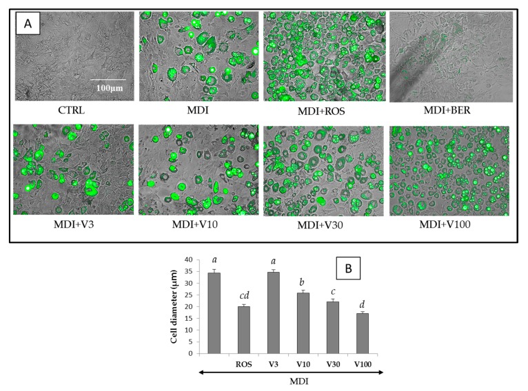
Figure 4.
Merged phase differences and fluorescent images and the diameter of differentiated 3T3-L1 cells on day 8 with reference compounds or vitexilactone of various concentrations. The 3T3-L1 cells were cultured in 24-well plates and differentiated with DMI mixture and each compound under the conditions described in the Materials and Methods section. Fluorescent staining of intracellular lipids was accomplished by adding BODIPY 493/503 to the medium. Undifferentiated cells, cells with the addition of MDI (a mixture of 0.5 mM 3-isobutyl-1-methyl xanthine (M), 0.1 μM dexamethasone (D), and 2 μM insulin (I)), rosiglitazone, berberine, and vitexilactone are indicated by CTRL, MDI, ROS, BER, and V, respectively. (A) Cell diameters were determined using ImageJ. Data are presented as the mean ± SD from 100 cells of three independent pictures. The same letters indicate that there are no differences between those groups, and different letters indicate significant differences (p < 0.05) (B).

