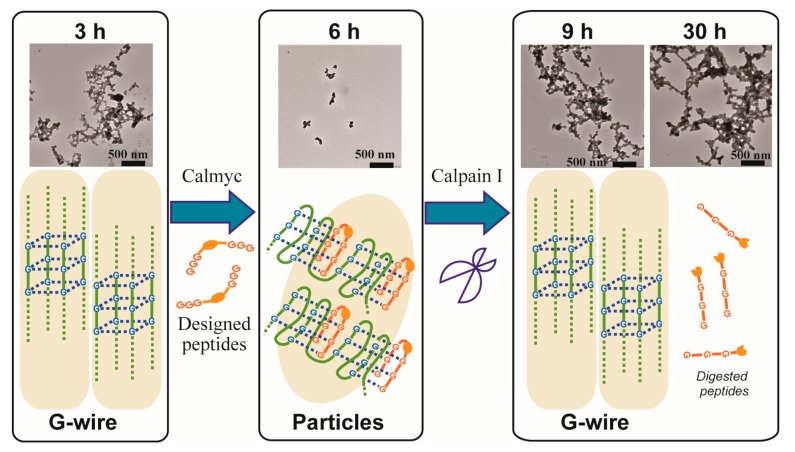
Figure 9.
TEM images of reversible switching of DNA nanowire formation with time using the peptide and the protease. MYC alone in the presence of Ca2+ was incubated for 3 h (the 3 h TEM image in the left column), the peptide was then added, and the sample was incubated for another 3 h (the 6 h TEM image in the middle column). After a total of 6 h of incubation, calpain I was added, and the sample was incubated for another 3 h or 24 h (the 9 h TEM image and the 30 h TEM image in the right column).

