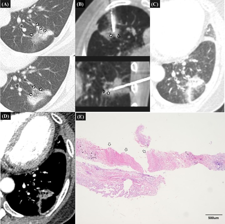
Fig 3. 52-year-old female patients without specific medical history, representative case of clinically significant hemoptysis due to penetrating injury of pulmonary artery and bronchiole after firing of biopsy gun.
A. Pre-procedural CT image shows a 32mm-sized sub solid mass in the left lower lobe with open bronchus sign (white arrow) which was suspected of primary lung cancer. B. Intra-procedural transverse and sagittal CT images before biopsy show that the introducer needle tip located within the mass. The bronchiole (white arrowhead) located just behind the introducer needle tip along the expected needle path of biopsy gun in transverse and sagittal CT images. The vessel (white arrow) which was seen in the medial margin of the mass lie along the expected track of biopsy gun in sagittal CT images. Biopsy was performed once and after the firing, hemoptysis began abruptly. C., D. Transverse enhanced CT image 20 minutes after the onset of hemoptysis shows the parenchymal hemorrhage along the introducer track. There was no evidence of extravasated contrast media around the mass. The patient was managed conservatively. E. The histopathologic examination of biopsy specimen shows pulmonary vessel larger than 1mm (white arrows) and small piece of bronchial epithelium. The pathologic diagnosis was consistent with primary lung adenocarcinoma.

