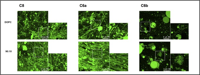
Fig 2. Confocal microscopy images of MLVs in presence of NBD labelled C8, C6a and C6b peptides.
MLVs are composed of DOPC/DOPG 90:10 M/M. Images were acquired on a laser scanning confocal microscope (LSM 510; Carl Zeiss MicroImaging) equipped with a plan Apo 63X, NA 1.4 oil immersion objective lens. For each field, both fluorescent and transmitted light images were acquired on separate photomultipliers and were analysed with Zeiss LSM 510 4.0 SP2 software.

