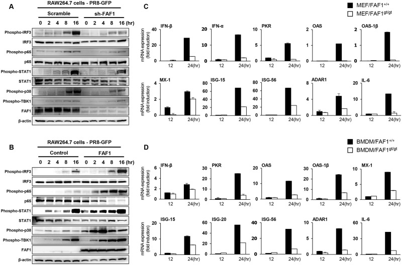
Fig 5. FAF1 activates the type I IFN signaling pathway and induces IFN-related gene expression.
(A and B) Control RAW264.7 (RAW-Scramble) and FAF1 knockdown RAW264.7 (RAW-sh-FAF1) cells (A) or control RAW264.7 (RAW-Control) and FAF1-overexpressing RAW264.7 (RAW-FAF1) cells (B) were infected with PR8-GFP (MOI = 2). At the indicated time points after infection, phosphorylated IRF3, p65, STAT1, p38 and TBK1, and total IRF3, p65 and STAT1 were measured in cell extracts by immunoblotting. β-actin was used to confirm equal loading of proteins. (C and D) Wild-type MEFs (MEF/FAF1+/+) and FAF1 knockdown MEFs (MEF/FAF1gt/gt) (C) and BMDMs isolated from FAF1+/+ (BMDM/FAF1+/+) and FAF1gt/gt (BMDM/FAF1gt/gt) mice (D) were infected with PR8-GFP (MOI = 1 and 3, respectively) for 12 hr, followed by total RNA extraction. Expression of mRNA encoding IFN-β, IFN-α, PKR, OAS, OAS-1β, MX-1, ISG-15, ISG-56, ADAR1 and IL-6 for MEFs and IFN-β, PKR, OAS, OAS-1β, MX-1, ISG-15, ISG-20, ISG-56, ADAR1 and IL-6 for BMDMs was analyzed by qRT-PCR. Data are presented as the mean ± SEM. Data are representative of at least two independent experiments.

