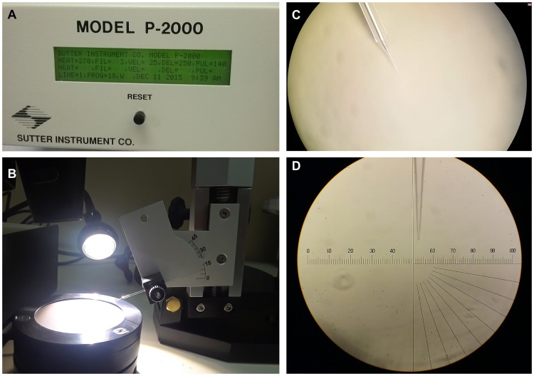
Fig 1. Needle preparation.
(A) Needles were pulled using a Sutter P-2000 micropipette puller. (B) (C) Pulled needles were beveled to an angle of 20° using a Sutter BV-10 Microelectrode Beveller, 104D fine abrasive plate. (D) Needles were inspected under a microscope to ensure the bore is not greater than 1 μm. NOTE: If there is no availability of a needle beveller, needles can be opened by breaking the bore towards a glass slide under the microscope (always verify the opening is smaller than 1 μm).

