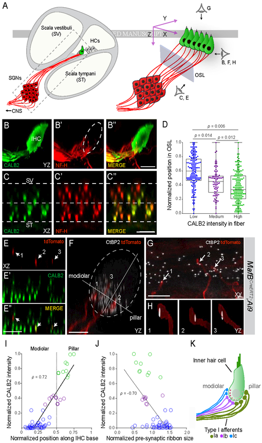
Figure 4: Type I SGN peripheral processes and synapses are anatomically segregated by subtype.
(A) Schematic depicting a cross-section of the cochlea (left) with a magnified view of the boxed area on the right. The three perspectives corresponding to the cochlear wholemount images in BH are indicated (right). Blue rectangle represents the plane of section through confocal image stacks of afferent fibers (red) extending through the osseous spiral lamina (OSL) to terminate along the basolateral surface of the hair cell (HC) (green). (B-C) Side (B) and cross-sectional (C) views of a wholemount cochlea stained for CALB2 (green, B,C) and NF-H (neurofilament heavy chain) (red, B’,C’), with merged images (B”,C”). CALB2+ fibers preferentially project towards the pillar side of the inner hair cell (IHC) compared to the total population of all NF-H+ SGN processes and are segregated along the scala vestibuli (SV)-scala tympani (ST) axis in the OSL (C-C”). CALB2 antibody also labels IHCs. (D) Quantification of afferent fiber distribution in the OSL. CALB2 fluorescent intensity levels were measured for all NF-H+ fibers in the OSL cross-section (n = 5 animals). Fibers were split into three groups based on CALB2 levels: ‘low CALB2’ (n = 165 fibers), ‘medium CALB2’ (n = 82 fibers), and ‘high CALB2’ (n = 174 fibers). Distance from the median center of each nerve bundle was calculated for individual fibers from each cluster. P values indicate results of Tukey’s HSD test following one-way ANOVA. (E-H) Individual tdTomato-labeled fibers (red) (E, E”) were traced in cochlear wholemounts from MafbCreERT2;Ai9 animals that were also stained for CALB2 (green, E’, E”) to assign subtype identity. Presynaptic ribbons were defined by co-staining for CtBP2 (white, F-H). In this example, three individual tdTomato-labeled SGN fibers (arrows) express ‘high’ (2), ‘medium’ (3), and ‘low’ (1) levels of CALB2 as they project through the OSL (E, E”). The same three fibers segregate along the modiolar-pillar axis of the IHC, shown in side view in F. Each tdTomato-labeled fiber terminates opposite a single presynaptic ribbon, shown in high resolution reconstructions (H). (I-J) Quantification of all analyzed fibers (n = 61, 5 animals) revealed that both fiber position (I; p = 0.72) and ribbon size (J; p = −0.70) correlate with CALB2 intensity. (K) Type Ia (green), Ib (purple), and Ic (blue) SGNs extend peripheral processes that are segregated in the OSL and along the modiolar-pillar axis of the IHC where they are apposed by presynaptic ribbons that decrease in size along the same axis. These features match those described for high, medium, and low SR SGNs. Scale bars: 10 μm (B, C, E, F); 5 μm (G). See also Fig. S5.

