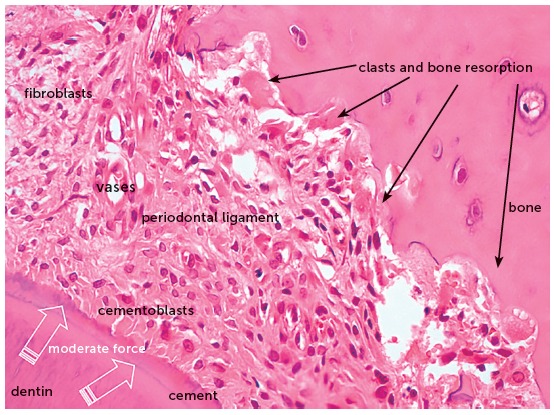
Figure 4. Microscopic aspects of rat molar root presented in Figure 3, at greater magnification, in axial section, after 4 days under moderate orthodontic forces. The image reveals the activity of clasts and other cells, thanks to the maintenance of periodontal structures, without hyalinization (HE, 40X magnification).

