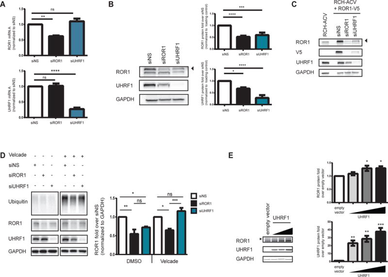
Figure 2. UHRF1 mediates ROR1 protein but not mRNA expression in RCH-ACV cells.

Cells were electroporated with siRNA and (A) 72 hours after electroporation, total RNA and whole protein lysates were harvested and tested for mRNA expression by qRT-PCR (N=3 **p=0.005, ****p<0.0001) and protein levels by (B) immunoblot (N=6 ****p<0.0001, ***p<0.0005, *p<0.05). mRNA and protein levels were normalized to a non-specific siRNA control (siNS). (C) RCH+ROR1-V5 cells were treated with siRNA and whole cell lysates were subjected to immunoblot (N=4). (D) RCH-ACV cells were treated with siRNA for 48 hours prior to treatment with DMSO or Velcade for 16 hours. Whole cell lysates were subjected to immunoblot. ROR1 protein was quantified and normalized to siNS samples treated with DMSO (*p<0.05, **p<0.005) or Velcade (*p<0.05, ***p<0.0005). UHRF1 protein was quantified and nomalized to siNS samples treated with DMSO (*p<0.05). Note that these data are from a contiguous blot (N=3). (E) HEK293T17 cells were transfected with either empty vector or a plasmid encoding UHRF1, and whole cell lysates were harvested 48 hours post-transfection. Levels of ROR1 (p<0.05) and UHRF1 (**p<0.005, ***p<0.0005) were detected by immunoblot. Error bars represent S.E.M. “ns” = not significant.
