



Abstract
Nuclear alterations are a hallmark of many types of cancers, including prostate cancer (PCa). Recent evidence shows that subvisual changes, ones that may not be visually perceptible to a pathologist, to the nucleus and its ultrastructural components can precede visual histopathological recognition of cancer. Alterations to nuclear features such as nuclear size and shape, texture, and spatial architecture reflect the complex molecular level changes that occur during oncogenesis. Quantitative nuclear morphometry, a field that uses computational approaches to identify and quantify malignancy-induced nuclear changes, can enable a detailed and objective analysis of the prostate cancer cell nucleus. Recent advances in machine learning-based approaches can now automatically mine data related to these changes to aid in diagnostic decision-making and prediction of PCa prognoses. In this review, we use prostate cancer as a case study to connect the molecular level mechanisms that underlie these nuclear changes to the machine learning computational approaches, bridging the gap between the clinical and computational understanding of PCa. First, we discuss recent developments to our understanding of molecular events that drive nuclear alterations in the context of prostate cancer: the role of the nuclear matrix and lamina in size and shape changes, the role of three-dimensional chromatin organization and epigenetic modifications in textural changes, and role the tumor microenvironment in altering nuclear spatial topology. We then discuss the advances in the applications of machine learning algorithms to automatically segment nuclei in prostate histopathological images, extract nuclear features to aid in diagnostic decision-making, and predict potential outcomes such as biochemical recurrence and survival. Lastly, we discuss the challenges and opportunities associated with translation of the quantitative nuclear morphometry methodology into the clinical space. Ultimately, accurate identification and quantification of nuclear alterations can contribute to the field of nucleomics and has applications for computationally driven precision oncologic patient care.
Keywords: Quantitative nuclear morphometry, Prostate cancer, Machine learning in medicine, Molecular-level nuclear changes, Nuclear architecture
1 | INTRODUCTION
Prior to the widespread utilization of prostate-specific antigen (PSA) in the late 1980s, of the patients that presented with prostate cancer (PCa), over 50% of them exhibited advanced disease (Veltri and Christudass, 2014). Now, however, use of the PSA test has resulted in overdiagnosis and the overtreatment of many indolent tumors that would not have caused harm during a patient’s lifetime. In addition, despite early interventions, nearly 20%-40% of patients still experience cancer recurrence following radical prostatectomy.
PCa biology can be characterized largely by genetic instability. While hereditary cancers only account for a minority of PCa, the majority of PCa cases emerge from somatic mutations. It is believed that the late onset of PCa suggests that genetic changes leading to oncogenesis accumulate over many years before clinical presentation (Knudsen and Vasioukhin, 2010). Studies indicate that chronic infections, inflammation, and numerous genotoxic insults can initiate genetic instability and lead to cancer formation through both genetic and epigenetic changes (Bianco-Miotto et al., 2010). Further, PCa is also considered to be a highly heterogeneous disease, which is reflected in the fact that multiple histologic patterns of cancer can be found within the same gland during Gleason scoring by expert pathologists. As such, due to the complexity and multitude of gene alterations that have been shown to drive PCa progression, finding a unifying genetic signature and identifying specific subtypes of PCa has proved elusive to date (You et al., 2016).
Transformation of normal prostate epithelium due to accumulated genetic and epigenetic changes can be phenotypically identified by observation of alterations to both the nuclear and tissue architecture (Jevtic and Levy, 2014). It has long been noted that disruptions to the organization of normal nuclear architecture is a hallmark of cancer (Zink et al., 2004). While defects in nuclear structure and function themselves do not cause cancer development, the altered nuclear organization in cancer cells is a manifestation of transformative and progressive oncogenic events (Madrazo et al., 2017). Malignancy-induced nuclear changes may have profound implications for three-dimensional chromatin organization and folding, the fidelity of genome replication, and the regulatory mechanisms of gene expression (Nickerson, 1998). Despite the many pleiotropic and pleiomorphic variations studied in the cancer cell nucleus, the underlying mechanisms that cause these changes and the functional links that lead to malignancy are poorly understood to date (Jevtic and Levy, 2014). Yet, changes to nuclear architecture, including nuclear enlargement and prominent nucleoli, are key features used by pathologists to identify cancerous regions from benign regions across many cancer types.
Recent efforts to quantify these changes to cancer cell nuclei using computer-aided methods for use in the diagnostic, prognostic, and therapeutic spheres have been substantial. Quantitative nuclear morphometry, a field that uses computational approaches to identify and quantify malignancy-induced nuclear changes, has emerged as a potential solution to meet the clinical needs of early identification of PCa at the cellular and molecular level. Semi-automated methods developed in the early 2000s showed initial benefits in the diagnostic space but required manual identification of cancer nuclei (Veltri et al., 2004). Previous limitations in efficiency and reproducibility have now sparked a revolution of new, fully automated algorithms that utilize machine learning to segment nuclei, extract and quantify nuclear features, and use these features to predict PCa progression. While several companion diagnostic genomic and proteomic assays are commercially available, they fail to capture the full extent of disease heterogeneity as they only sample a small part of the tumor landscape. On the contrary, computer-assisted approaches are able to identify subvisual nuclear changes in histological images that are unavailable at the level of the human eye nor in genomic and proteomic analyses and allow for a larger coverage of tumor landscape in both cancerous and benign-adjacent regions. Machine learning methods are able to uniquely discover patterns and relationships within and between cancer cell nuclei from complex datasets to effectively predict future outcomes (Kourou et al., 2015). This digital transformation in oncology and pathology has the potential to provide new tools to aid clinicians in decision-making for more precision-based approaches (Bhargava and Madabhushi, 2016).
We identify a gap in the current literature: while research discovering the molecular mechanisms driving nuclear structural changes and research pioneering the computational algorithms for quantitative nuclear morphometry are both individually well-developed, there seems to be a disconnect between the molecular mechanisms and the morphometric changes. We believe that much of the existing literature on the computational approaches often neglect discussions of the molecular events behind the nuclear features that the algorithms can now identify. Thus, this review is motivated toward molecular biologists and oncologists who may be unfamiliar with the computational algorithms that are currently being applied to optimize PCa care, thus bridging the molecular events with the computational algorithms. We will use PCa as a case study and will expand on the potential to utilize these approaches to serve as a new “biomarker” for predicting outcome-related oncological metrics, such as tumor aggressiveness, tumor recurrence, and patient survival. We define the scope of this review to only include recent advances (2011-present) in quantitative nuclear morphometry in the PCa setting as previous reviews discuss the early history and developments of these methods (Veltri et al., 2012). Studies describing computational applications of prostate gland morphometry and nuclear morphometry in other tumor types, while important, have been excluded from this analysis. In Section 2, we introduce the nuclear features that can be characterized by current quantitative algorithms and describe recent developments regarding the molecular mechanisms governing these changes in the context of PCa. In Section 3, we will then connect these molecular level changes with the new computational approaches designed to automatically identify nuclei from histological images, extract nuclear features, and use these features to predict PCa-related outcomes. In Section 4, we highlight challenges and opportunities related to the clinical application of these approaches and the potential of nucleomic data to supplement existing clinical metrics to aid clinicians for precision oncologic care.
2 | CANCEROUS MOLECULAR LEVEL CHANGES THAT DRIVE NUCLEAR MORPHOMETRIC FEATURES
Normal nuclear function is tightly regulated and compartmentalized across many cell types. Among the chief functions of the nucleus are to organize chromatin and regulate gene expression (Jevtic et al., 2014). Consequently, accumulated genetic insults often result in malignancy-induced changes to the nucleus, which are among the most remarkable changes in cells during cancer progression (Nickerson, 1998). These “subvisual” changes often precede the histopathological recognition of cancer (Leman and Getzenberg, 2008). In the context of quantitative nuclear morphometry in prostate cancer, features that are extracted to characterize these nuclear architecture and structural changes in cancer are typically divided into three broad domains: measures of nuclear size and shape, measures of nuclear textural changes, and measures of the spatial topology of the surrounding microenvironment (Bhargava and Madabhushi, 2016). Algorithms extract both handcrafted and unsupervised features: handcrafted features refer to those which can be attributed to the intuitive measurements in the image (e.g. nuclear shape or texture) while unsupervised features refer to machine-derived relationships that are learned following training of the algorithm from the initial set of tissue specimen images. In theory, these algorithms capture the phenotypic result of the complex molecular events that undergo significant changes during oncogenesis and tumor progression. These molecular-level events have important morphological ramifications for the nuclear architecture, structure and function, both within the nucleus and within the local tissue microenvironment. As such, in this section, we highlight the recent advances in the understanding of the underlying mechanisms that dictate nuclear size and shape, texture, and spatial microenvironment at the molecular level that machine learning algorithms are able to identify at the morphometric level. Figure 1 depicts the important nuclear substructures that give rise to the detected features summarized in Table 1.
Figure 1.
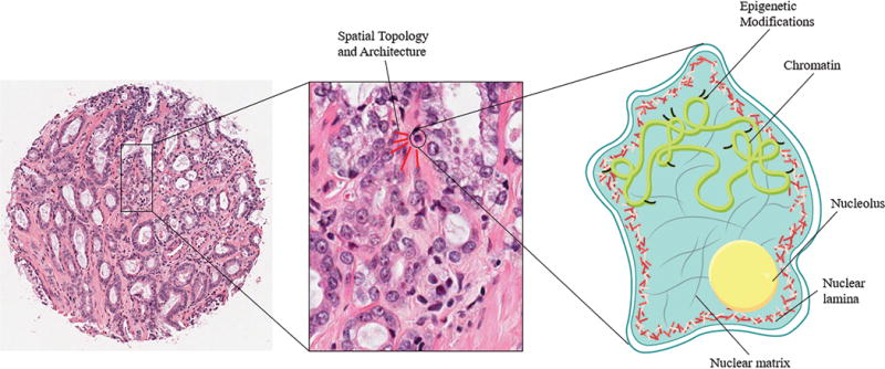
The nucleus can be a critical component of the visual diagnosis of PCa. Subvisual structural and architectural alterations can precede visible histopathological changes. As such, we identify and implicate the ultrastructure and microenvironment for the subvisual nuclear changes that occur during cancer progression, which can ultimately be identified by quantitative nuclear morphometric analysis for the identification and prediction of PCa. These components include: (1) The nuclear lamina and matrix, which undergo significant expression and conformation changes during oncogenesis and can be responsible for driving nuclear size and shape irregularities; (2) Three-dimensional chromatin organization and epigenetic changes, which drive textural changes to the nucleus; and (3) Spatial topological and architectural arrangement patterns, which can be characteristic of pre-malignant changes.
Table 1.
Common features that are extracted from prostate histological images using machine learning algorithms to describe nuclear size and shape, texture, and topological and architectural changes.
| (A) Size & Shape Features | |||
|---|---|---|---|
| Feature Subtype | Handcrafted or Unsupervised | Specific Features Extracted from Nuclei | Relevance to Prostate Histopathology |
| Nucleus Morphology (2D) | Handcrafted | Area, Perimeter, Elliptical major & minor axis lengths, Overlap ratio, Diameter/Radius, Number of nucleoli, Nucleolar min/max diameter, Bounding box, Circularity, Eccentricity, Bounding circle ratio, Distortion, Roundness | Characterize nuclear pleomorphism through descriptors of size, shape, and boundary |
| Nucleus Morphology (3D) | Handcrafted | Object volume, Volume of 3D bounding box, Diameter of sphere with same volume as bounding box, ratio of the object volume to the bounding box volume | 3D characterization enables additional volume measurements |
| (B) Texture Features | |||
|---|---|---|---|
| Feature Subtype | Handcrafted or Unsupervised | Specific Features Extracted from Nuclei | Relevance to Prostate Histopathology |
| DNA Content Features | Handcrafted | Absorbance (Mean, Median, SD, Min/Max), DNA ploidy, DNA index | DNA ploidy related to staining where amount of color developed is proportional to the amount of DNA |
| Chromatin-Related Texture | Handcrafted | Transmission, Intensity, Cluster prominence, Homogeneity/Heterogeneity, Entropy, Luminance, Optical Density, Cluster shading | New algorithms can enable pixel-level analysis of textural changes that show the spatial distribution of chromatin in the nucleus |
| Curvelet-Based Features | Unsupervised | Variance, Energy, Entropy, Skewness, Kurtosis | Curvelet-based features are used to define the boundaries of curvilinear objects, such as nuclei |
| Intensity-Derived Features | Unsupervised | Haralick features, Gabor features, Local binary patterns | These image-based features are used for a pixel-level texture representation for an optimized analysis of texture changes in the nucleus |
| Nucleotyping (Hveem et al., 2016) | Handcrafted | Grey-level entropy matrix | Represents the spatial variation of DNA density and three-dimensional euchromatin/heterochromatin organization |
| (C) Topological & Architectural Features | |||
|---|---|---|---|
| Feature Subtype | Handcrafted or Unsupervised | Specific Features Extracted from Nuclei | Relevance to Prostate Histopathology |
| Nuclear Architecture | Handcrafted | Nucleus centroids as nodes in a graph | Enables identification of nuclear clusters to create a local graphical analysis of nuclear architecture |
| Voronoi Diagram | Unsupervised | Area (Average, SD), Area & Perimeter disorder | Global measure of nuclear architecture characterizing the spatial interactions between nuclei |
| Delaunay Triangulation | Unsupervised | Triangle area (Average, SD), Side length (Min/Max, Average, SD) | Identify global triangle-based spatial relationships of nuclei |
| Minimum Spanning Tree | Unsupervised | Edge length (Average, SD, Min/Max, Disorder) | Enables spatial topological analysis by vertices (representing nuclei) in a tree-like manner |
| Cell Cluster Graph | Unsupervised | Eccentricity, Clustering, Isolated nuclei, Connected nuclei, Skewness of edges | Graph-theory based approach that defines a graph node as a nuclear cluster rather than an individual nucleus to enable spatial relationships |
| Geometric Descriptors | Unsupervised | Global fractal dimensions, Shannon entropy, Lacunarity | Characterize the spatial geometry of PCa cells and nuclei by measuring distance from a nucleus to its closest neighbor, disorder, and degree of gaps between cells and nuclei |
| Cell Orientation Entropy (Lee et al., 2013) | Unsupervised | Entropy, Energy, Correlation, Contrast (Variance) | Orientation entropy is a model of disorder of nuclear orientation within local neighborhoods of specimen images |
2.1 | Nuclear Size and Shape Alterations in Cancer
Measurements of nuclear size and shape and their derivatives are among the most common features extracted during quantitative nuclear morphometric analysis. Table 1A shows relevant size- and shape-related features that have been used in quantitative nuclear morphometry for PCa. Many factors have been linked to altered nuclear size and shape in cancer, including ploidy, changes to lamin expression, alterations to chromatin organization, nuclear envelope changes, and deregulated nuclear matrix-associated protein composition (Dey, 2010). Nuclear dysmorphia is a nearly ubiquitous feature of cancer and provides further motivation for studying the main structural components of the nucleus: the nuclear matrix and nuclear lamina (Irianto et al., 2016). While many changes do in fact occur in the cancer cell nucleus, the underlying ultrastructural changes orchestrated by the nuclear matrix and nuclear lamina strongly contribute to the observed physical size and shape changes.
The nuclear matrix is widely considered to be the main scaffolding of the nucleus, consisting largely of hundreds of proteins interconnected with the ribonucleoprotein network and nuclear lamina (Nickerson, 1998). Notably, the nuclear matrix plays a significant role in the spatial organization of chromatin through infrequent yet highly specific matrix-attachment regions (MARs) (Zink et al., 2004). MARs are typically characterized by AT-rich DNA sequence attachment points and in conjunction with the nuclear matrix, arrange chromatin into loops. Additionally, MARs have been shown to have a modulation role in critical functions such as RNA splicing, DNA replication, and gene expression by serving as an anchoring point for catalytic and regulatory factors to mediate transcription- and post-transcription-related functions (Nickerson, 1998).
In PCa tumor progression, the nuclear matrix protein composition and chromatin organization is altered due to changes in the MAR loop domains (Barboro et al., 2012). In a recent study, DNA loop organization was studied in the PCa cell lines LNCaP, considered a less aggressive PCa cell line, and PC3, considered a more aggressive PCa cell line. The nuclear matrix was well organized in the LNCaP cell line with many nuclear matrix proteins bound to MAR sequences, creating short DNA loop domains with many attachments. However, in the more aggressive PC3 cells, irregularities were observed in the nuclear matrix, with fewer proteins attached to MAR sequences, creating large loop domains (Barboro et al., 2012). Deregulation of the nuclear matrix allowed for critical chromatin unfolding in the aggressive phenotype, and the previously tightly bound heterochromatin was hence decondensed from the periphery to a more interior localization. While there are many proteins associated with the nuclear matrix, only a small subset of specific MAR-binding proteins seems to significantly affect chromatin reorganization during carcinogenesis. SATB1 is a well-established MAR-binding protein that has been associated with the aggressive PCa phenotype. SATB1-coordinated changes to MAR-nuclear matrix interactions could lead to observed variations in loop domains, thus altering gene expression and triggering a more aggressive PCa phenotype (Mao et al., 2016).
The nuclear lamina is a dense network of intermediate filaments that line the inner nuclear membrane while providing mechanical strength to the nucleus, organizing chromatin, and modulating DNA transcription. Mutations to the lamin genes LMNA and LMNB result in diseases collectively termed laminopathies, which are known to show abnormal nuclei with herniations and lobulations (Saarinen et al., 2015). Altered lamin expression is also considered to be an important step involved in the malignant transformation (Khan et al., 2018). Lamins directly interact with chromatin through lamin-associated domains (LADs) and are major determiners of chromatin positioning in the nucleus. LADs cover more than a third of the human genome, making it one of the most prominent epigenetic features in the nucleus. Both A-type and B-type lamins expression abnormalities have been shown to play a role in PCa progression. Lamin A regulates gene expression by binding chromatin at the nuclear periphery, which is typically a transcriptionally repressive region (Sakthivel and Sehgal, 2016; Lever and Sheer, 2009). Further, lamin B1 was shown to correlate with biochemical recurrence and local spread within the prostate gland, while an inverse relationship was observed between lamin A/C levels and nuclear deformability (Saarinen et al., 2015). Recent evidence implicates lamin A/C as the primary director of nuclear stiffness (Davidson and Lammerding, 2014). Low levels of lamin A/C could impact the metastatic potential of PCa by enhancing cell migratory ability by promoting lymphovascular invasion and an increased risk of lymph node metastasis contributing to an unfavorable disease-specific survival.
2.2 | Nuclear Textural Changes and Measures of Disorder
Both early and recent quantitative nuclear morphometric approaches employ measures of nuclear texture to evaluate changes to the 3D chromatin organization, changes to the quantity of genetic material, and changes to the global epigenetic landscape (Veltri et al., 2012; Hveem et al., 2016). Table 1B highlights the many features that have been used to characterize these changes. Genetic and epigenetic variations are thought to drive large-scale nuclear alterations during carcinogenesis and cancer progression, even though the exact mechanisms have yet to be delineated. Recent evidence further posits that genetics and epigenetics behave in a reticulate manner – it is clear that alterations in epigenetic factors can lead to genetic mutations; similarly, genetic mutations of epigenetic regulators can lead to an altered epigenome (You and Jones, 2012). While cancer is characterized as a disease initiated and driven by somatic genome aberrations, discovering how these genomic and epigenomic alterations culminate in malignant transformation must precede the development of targeted individual therapeutics. In many cases, textural changes in histopathological specimens result from variations in staining intensity using hematoxylin & eosin (H&E) and DNA Feulgen stains. Both stains can result in textural changes to the nucleus: the basicity in the H&E stain selectively targets the DNA in the nucleus, while the Schiff-base reaction in the Feulgen stain results in identification of chromosomal material. Algorithms now have the ability to detect minute differences in staining across nuclei reflecting the complex genetic and epigenetic interplay during oncogenesis.
Ionizing energy deposits (e.g., x-rays and UV light), reactive oxygen species, chemical mutagens, and replication errors are among the most common lesion-causing DNA agents in PCa carcinogenesis (Mateo et al., 2017). These exogenous and endogenous stresses illicit distinct repair mechanisms to mediate the restoration of DNA strand breaks and mismatches. If a malfunction occurs but apoptosis does not ensue, joining of different genomic regions could produce rearrangements that may preclude carcinogenesis (Lever and Sheer, 2009). In PCa, common genetic alterations include mutations, gene fusions, copy number variations, and translocations, often occurring simultaneously over the course of many years (Veltri and Christudass, 2014). There is a substantial heterogeneity amongst genetic aberrations observed in PCa as evidenced by the wide spectrum of the molecular abnormalities and unpredictable clinical course (Abeshouse et al., 2015). This heterogeneity makes subtyping PCa for treatment stratification difficult. However, in a recent study, The Cancer Genome Atlas (TCGA) initiative showed that 74% of tumors fell within one of seven identified subtypes based on common gene fusions. Notably, multiple studies have identified ERG gene fusions as drivers of PCa; one group showed that multiple distinct ERG fusions existed within a single cancer nodule from a patient sample (Abeshouse et al., 2015; Cooper et al., 2015). Recent advances in genomics have also allowed for the identification of frequently mutated genes in PCa, including SPOP, TP53, FOXA1, and PTEN.
Epigenetic modifications seem to play an ever-increasing role in the development of PCa through altered gene expression. With the late onset of PCa, it is believed that lifestyle factors such as diet, stress, and levels of physical activity can gradually alter both the genome and epigenome during a patient’s lifetime. As such, epigenetic marks play a key role in the control of gene activity. DNA hypomethylation has been linked to activation of oncogenes, and signs of chromatin histone modifications have shown to have a predictive ability in characterizing prostate tumor grade and recurrence potential (Seligson et al., 2005). Recent immunohistochemical analysis in a 279-patient PCa cohort revealed that high levels of histone modifiers H3 lysine-18 acetylation (H3K18Ac) and H3 lysine-4 dimethyl (H3K4diMe) were predictors of relapse-free survival (Bianco-Miotto et al., 2010). While exact mechanisms or implications of histone modification levels are not yet well understood in the context of PCa, elevated or depressed histone levels may occur concomitantly with tumor progression, resulting in abnormal gene expression patterns. Another study showed the significance of changes in DNA methylation status to the genes GSTP1, APC, and RASSF1 in high-grade PCa (Van Neste et al., 2017). Epigenetic hypermethylation of these genes, which is associated with DNA detoxification, apoptosis, and cell cycle regulation, had a silencing effect in the context of PCa, and using this epigenetic signature for prognostic purposes revealed a role for identifying high-risk PCa.
Modifications at the genetic level (DNA damages and their structural consequences) and epigenetic level have implications for the establishment of cancer cell nuclei organization and architecture (Naimark et al., 2016). These multilevel changes are nearly always associated with changes to chromatin condensation or location. Most significantly, the structure and spatial organization of the genome has physical constraints, such as those related to the helical turning of one strand around the other. Further constraints are associated with DNA supercoiling around nucleosomes, which govern chromatin compaction. Strand breaks can cause a relaxation of the constraints, shifting the chromatin into a more disordered conformation and subsequently altering nuclear properties. Further, epigenetic modifications such as methylation, phosphorylation, and acetylation of nuclear structural components such as lamins can also dictate nuclear organizational changes, showing the interconnectedness between possible textural changes and size and shape changes.
2.3 | Spatial Topology and Influence of the Tumor Microenvironment
While intranuclear processes serve as the molecular underpinnings of cancer-induced alterations to the nuclear architecture, the role of the microenvironment cannot be understated. In PCa, it is now becoming clear that a detailed spatial analysis of the nuclear orientation in the context of the local microenvironment can yield important diagnostic and prognostic subtleties. Advanced quantitative nuclear morphometric methods now have the capability to identify pre-cancerous changes in adjacent normal-appearing benign nuclei as well as spatial relationships between regional cells and nuclei that could have implications for tumor aggressiveness and recurrence. New evidence suggests that biochemical and mechanical cues from other cells, the extracellular matrix, and the cytoskeleton can all impact nuclear mechanics, genome organization, and transcription (Uhler and Shivashankar, 2017), further complicating the mechanisms behind the architectural changes. Evidence of a field cancerization in PCa has also been widely shown, providing additional support for the influence of the microenvironment in determining nuclear properties and potential for metastasis during cancer progression (Veltri et al., 2004; Verdone et al., 2015; Lee et al., 2017).
When cells are subjected to extreme mechanical deformations, such as shear, tensile, or compressive forces, alterations to gene programs occur as a counterbalance to such physiomechanical stresses. In extreme circumstances, these local forces can be influential enough to induce transdifferentiation or a disease-triggered state (Uhler and Shivashankar, 2017). Continuity between the extracellular matrix, the cytoskeletal network, and chromatin provide a direct mechanotransduction bridge for the extracellular environment to alter genome organization and nuclear properties (Irianto et al., 2016). Specifically, linker of nucleoskeleton and cytoskeleton (LINC) complexes allow the direct transmission of extracellular mechanical signals. Perinuclear actin-mediated connections exert mechanical strains on the nucleus, influencing nuclear morphology (Kim and Wirtz, 2015). LINC attachments to the nuclear lamina can contribute to spatiotemporal changes in chromosome condensation and expression. Extracellular mechanical forces can also initiate biochemical pathways that effect nuclear dynamics through tuning of post-translational histone modifications. While extracellular mechanical forces and biochemical pathways seem to play a significant role in disease onset and potential metastatic capabilities, its role as a director of cancer or a downstream effector of altering the nuclear architecture to induce metastasis is still unknown.
The “field cancerization” effect in prostate carcinogenesis has been well established. The field effect can be characterized by abnormal genetic, epigenetic, and protein molecular changes to regions outside of the main tumor foci that are associated with cancer (Risk et al., 2010). Additionally, alterations to benign nuclei are also seen in areas adjacent to the main tumor, thus allowing for nuclear architectural characterization of the field effect. In prostate cancer, one of the only remaining solid tumors diagnosed solely from random needle biopsy, sampling errors have been attributed to missing tumor foci during biopsy altogether or under-diagnosis from missing the main tumor foci. However, even with a poor biopsy sample, characterization and quantification of benign samples have shown to provide prognostic information. Contemporary imaging and quantification modalities have allowed for a greater understanding of the field effect, which now encompasses a wide range of molecular-level changes in histologically “normal” samples (Nonn et al., 2009). In the study cited above that characterized a three-gene signature (GSTP1, APC, and RASSF1) for identifying epigenetic methylation changes in PCa, methylation patterns in adjacent normal-appearing cells were also associated with the presence of latent PCa (Van Neste et al., 2017). In addition to revealing epigenetic changes in tumor tissue samples, a predictive model captured the epigenetic field effect to stratify patients at the time of biopsy. Various recent studies have also confirmed architectural changes in normal-appearing adjacent nuclei. One such study employed a novel imaging modality that measured nanoscale architectural alterations in normal epithelial cells and was able to discriminate early stages of PCa (Roy et al., 2015). Despite these demonstrated changes, the molecular foundation of the field effect remains ill-defined to date.
3 | ADVANCES IN ALGORITHMS FOR QUANTITATIVE NUCLEAR MORPHOMETRY
Visual and subvisual characteristics embedded in histopathological images contain diagnostic and prognostic properties that reveal the manifestations of underlying molecular foundations of disease. In the PCa setting, clinicians have relied on subjective assessments of histopathological specimens obtained from biopsy and surgery to inform disease management and treatment decisions. PCa is known to have a high glandular and nuclear heterogeneity even within different regions of the same tumor. Even among specialists, discrepancies in determining tumor grade are nontrivial. Manual grading remains a laborious process, often resulting in poor score reproducibility between pathologists (Hamilton et al., 2014). Computer-assisted quantitative approaches can ameliorate many of these challenges by providing an objective algorithm that extracts even the most minute differences unapparent to the human eye. In turn, extraction of large amounts of nuclear data can supplement other data and contribute to a precise diagnosis and prediction of disease course (Djuric et al., 2017). While semi-automated quantitative nuclear morphometric approaches of the early 2000s still required significant time and expertise to perform reproducibly, recent advances in computer science, image analysis, and scanning microscopy have revolutionized automated machine learning algorithms which can identify and quantify nuclear morphology in a high-throughput manner. Machine learning methods “learn” from a set of data samples by extracting patterns and relationships to automatically identify features which will be able to classify those samples. In quantitative nuclear morphometry, applications of image processing and machine and deep learning can be used to perform tasks such as nuclear segmentation, feature extraction for diagnostic decision-making, and prognostication of PCa-related outcomes. The recent (2011-present) studies and advances within the field of quantitative nuclear morphometry in PCa are summarized in Table 2. While literature covering computational applications to gland morphology and nuclear morphometry for other types of solid tumors exists, this section will focus specifically on quantitative nuclear morphometry for PCa.
Table 2.
Summary of the recent advances (2011-present) in the algorithmic approach to nuclear segmentation, feature extraction, and prognostication of PCa-related outcomes from histopathological images.
| (A) Nuclear Segmentation | ||
|---|---|---|
| Study | Algorithms Used in Analysis | Major Advancement |
| Ali et al., 2011 | Used watershed-based image initialization with subsequent level set formulation for segmentation | Improved computational efficiency of shape-based segmentation method by selective invocation of the active contour scheme |
| Kalinin et al., 2015 | Used publicly available FARSIGHT toolkit for nuclear segmentation with post-segmentation hole filling and filtering; used Trainable Weka Segmentation for intra-nuclear objects | Successfully segmented nuclei and nucleoli using three-dimensional methods; created a publicly available dataset of segmented nuclear images |
| Gertych et al., 2016 | Used 3D radial symmetry transform followed by an adaptive post-processing | Achieved a true positive segmentation rate of 83.3% |
| Zarei et al., 2016 | Used a Gaussian filter, RGB to grayscale conversion, and gradient-weighted thresholding for segmentation | Obtained a 96.6% true positive segmentation rate with a 12% false positive rate |
| (B) Feature Extraction for Diagnostic Decision Making | ||
|---|---|---|
| Study | Algorithms Used in Analysis | Major Advancement & Result |
| Doyle et al., 2011 | Utilized a class-balanced active learning classifier to address the large amount of non-target class exemplars (i.e. benign tissue) are included in the training data | Achieved 2% greater accuracy and 3% greater AUC than other comparative classifiers |
| Doyle et al., 2012 (BMC Bioinformatics) | Used a cascaded approach, where algorithm makes successive binary classifications to classify similar specimens together | Extraction of architecture- and texture-based features resulted in a positive predictive value of 0.86 in classifying 2000 tissue images |
| Doyle et al., 2012 (IEEE Trans Biomed Eng) | Boosted Bayesian multiresolution to analyze successive image resolutions to generate a spatial analysis of the specimen | Achieved superior results over a random forest and individual feature classifier with an eightfold savings in computational time |
| Gann et al., 2013 | Used K-means clustering and watershed for segmentation and technician to manually select regions of interest for analysis | Multifeature nuclear morphometric score incorporated 5 of the most significant nuclear features from the original set of 52 to discriminate cancer from benign prostate cells with an AUC = 0.91 in the training set and 0.79 in the validation set |
| Verdone et al., 2015 | Used commercially-available image processing software to extract features; employed k-nearest neighbor, SVM, and multinomial logistic regression to incorporate nuclear features for discrimination | Showed that the epithelial-to-mesenchymal transition in PCa is associated with changes to the nuclear structure; achieved AUC of 0.95 using SVM and 0.96 using multinomial logistic regression to separate PC3-Epi from PC3-EMT cells |
| Gertych et al., 2015 | Used a two-step SVM and RF classification scheme to stratify stromal, benign, and cancerous tissue | Extraction of intensity-based staining features allowed for a pixel-based classification and successfully identified PCa regions from stromal and benign regions |
| Lin et al., 2016 | Employed a four-class tree classifier with two sequential SVMs trained with curvelet-based texture features | First SVM successfully identified homogenous tissue (GS 3+3 and GS 4+4) from heterogenous tissue; second SVM trained to distinguish between GS 3+4 and GS 4+3; overall scheme achieved a 93.75% testing accuracy |
| Waliszewski, 2016 | Attempted to validate previously reported classes of equivalence based on fractal geometrical models to characterize the spatial distribution of PCa cell nuclei for Gleason grading | Novel quantitative criteria based on the spatial complexity and spatial diversity features allowed for separation of low and highly aggressive PCa cases |
| Kwak and Hewitt, 2017 | Employed an automated, cooperative, multi-view boosting algorithm to characterize prostate tissues at different resolutions | Achieved an AUC of 0.98 for detecting cancer from non-cancer |
| Wang et al., 2017 | Utilized a novel discriminative feature scaling scheme to find and combine the optimal image resolutions at which the most distinguishing features could be identified | Achieved a 66% decrease in computational times compared to a scheme extracting features from all scales while achieving a comparable classifier performance to detect cancer regions on a per pixel basis |
| (C) Prognostication of PCa-Related Outcomes | ||
|---|---|---|
| Study | Algorithms Used in Analysis | Outcome Measured & Result |
| Ali et al., 2013 | Cell Cluster Graph to characterize local spatial architecture; trained a SVM classifier | Biochemical recurrence; methodology correctly classified 83.1% of recurrent cases from an 80-patient TMA |
| Lee et al., 2013 | Used cell orientation entropy to quantitatively model disorder of nuclear orientations; utilized RF classifier | Biochemical recurrence; cell/nuclear orientation features achieved an accuracy of 82.7% to classify recurrent cases from a 39-patient dataset |
| Hveem et al., 2016 | Developed a novel marker of epigenetic aberrations (Nucleotyping); used algorithm to derive grey-level entropy matrix values at various radial positions in the nucleus | Recurrence after radical prostatectomy; Nucleotyping predicted recurrence with a HR of 3.3 |
| Lee et al., 2016 | Identified the most predictive of recurrence based on nuclear shape and architecture from cancerous and benign-adjacent regions were evaluated using a RF classifier | Biochemical recurrence following radical prostatectomy; Tumor-adjacent benign field nuclear arrangement and shape descriptors predicted recurrence with an AUC = 0.77 |
| Kleppe et al., 2018 | Attempted to validate the results from Hveem et al., 2016 in a pan-cancer study | Cancer-specific survival; in prostate carcinomas, Nucleotyping was not significantly prognostic (HR = 1.7) but showed promising results |
3.1 | Nuclear Segmentation
Following biopsy, tissue specimen slides are prepared for image analysis. Prior to the extraction of pertinent features that facilitate cancer detection and diagnosis and prognostication of disease progression, image processing algorithms must accurately and robustly delineate nuclei in images with little need for human intervention. In whole slide images, segmenting or identifying nuclei in the cell type of interest (namely epithelial cells) from both background artifact and from other cell-type nuclei is a front-end problem that can greatly impair accurate downstream feature extraction and model predictions. Variations in nuclear shapes, overlapping and close-contacting nuclei, chromatin texture, and staining intensities of background artifacts can all impede identification of individual nuclei on a slide level (Gertych et al., 2016). However, a computationally expensive segmentation scheme that accounts for all of this variability can result in high processing times prior to feature extraction or disease outcome prediction. Advances in segmentation schemes attempt to enhance accuracy while reducing computational overhead. Ultimately, the nuclear segmentation scheme, as depicted in Figure 2A, seeks to establish accurate processing steps to produce an efficient yet accurate nuclear boundary to make image data more suitable for downstream feature extraction.
Figure 2.

Quantitative nuclear morphometric involves three critical steps: nuclear segmentation, nuclear morphometric feature extraction, and prognostication of PCa-related outcomes. (A) proper segmentation of nuclei from benign nuclei and background features relies on novel segmentation schemes that resolve nuclear overlapping and poor staining contrast. [Image adapted from Ali et al., 2011 with permission]. (B) Feature extraction can include nuclear size and shape features, textural features, or spatial topographical features. Left panel: depicts number of neighboring epithelial nuclei; center panel: depicts categorization of nuclear shape contexts; right panel: depicts distance fractal dimensions. [Image adapted from Kwak and Hewitt, 2017 with permission]. (C) Quantitative algorithms that utilize a number of different machine learning classifiers can use these features to create models to distinguish cancer from non-cancer, identify aggressive cancer phenotypes, or predict important PCa-related outcomes such as biochemical recurrence, progression, or mortality. Image depicts utilization of nuclear features to discriminate a more advanced PCa phenotype. [Image adapted from Carleton et al., 2018 with permission].
In a 2011 study, Ali et al. described a novel adaptive active contour segmentation scheme, termed AdACM, that relied on shape based active contours to resolve overlapping nuclei in prostate histopathological images. In TMA images, it is common for nuclei to be overlapping or confluent in regions, thus making overlap resolution a necessary aspect of a segmentation algorithm. In a shape-based segmentation scheme, a “shape prior” term is associated with the contours that identify nuclear boundaries. To obtain rough delineations of nuclear boundaries, the authors employed a watershed initialization, which relies on differences in intensities within an image to create rough topographical lines that outline objects of interest from the background, creating a hypothetical map on the image. Using this intensity-based initialization as a base for rough nuclei outlines, concavity points were calculated using three consecutive points around the nuclei borders, a concept first proposed by Fatakdawala et al. in 2010. High amounts of concavity points are characteristic of contours that enclose multiple objects, thus allowing for the identification of overlapping nuclei. Depending on the number of concavity points detected, the algorithm can identify non-overlapping nuclei against overlapping nuclei. While shape-based methods had been previously studied for object segmentation, the authors uniquely selectively invoke the shape-based term only when “prior” nuclei were found to have high concavity points predicting overlapping regions. As the shape-prior term is associated with high computational costs, selectively invoking the term only when necessary to resolve overlaps is critical to keeping reasonable image processing times. Quantitative analysis showed that the AdACM model achieved a nearly 90% success rate in segmenting overlapping nuclei. Further, compared to an active contour model that did not selectively invoke the shape prior term, the novel AdACM algorithm resulted in a greater than 100% more efficient segmentation scheme.
Even despite computational savings from previous methods, the active contour model with selective invocation of shape prior terms showed relatively large processing times for relatively small image sizes (250 seconds for a 200x200 pixel image). A 2016 study on Feulgen-stained tissue microarrays (TMAs) from 33 PCa patients employed a multistep mean gradient intensity thresholding algorithm to localize nuclei (Zarei et al., 2016). After employing an initial Gaussian filtering method to remove noise and small debris elements, a mean gradient thresholding approach followed by a Hough transform was used to further delineate nuclei. While the authors reported an initial complication with identifying overlapping nuclei, the Hough transform fits an elliptical shape to edge of the nuclei around the detected overlapping objects. In comparison to the AdACM method presented above, this method processed 1024×1024 pixel images in about 120 seconds with a 96.6% true-positive rate, showing further improvement upon the 250 seconds reported by Ali et al.
While two-dimensional segmentation algorithms are well-established, there has been a recent interest in high-resolution three-dimensional (3D) algorithms, which can be largely grouped into Watershed-based, hybrid, or deformable model-based methods. Introducing the additional dimension for segmentation purposes can result in significant functional information for nuclear architectural changes in cancer but can also result in inefficiencies with respect to image intensity variability and cell confluency (Gertych et al., 2016). A novel 3D-radial symmetry algorithm has been proposed, which produces a transformed image based on the circularity of nuclei that can then be subsequently analyzed using a Watershed-based method (Gertych et al., 2016). The 3D-radial symmetry preprocessing method yields a signal that is converted to large seeds fitted into the boundary of the binary nuclear mask. Further, using 3D features in combination with traditional 2D features can improve segmentation performance as shown in the PCa PC3 cell line (Kalinin et al., 2015). A gradient boosting supervised classifier was used as the optimal methodology for segmentation using 2D and 3D features.
Computational efficiency is a chief concern for the nuclei segmentation methodologies, especially when segmentation is but the first step in a fully automated machine learning algorithm that ultimately aims to have diagnostic and/or prognostic potential. Compromising accuracy of nuclei segmentation to reduce computational costs can result in downstream model errors. Building on previous work, new approaches have employed deep learning algorithms to implicitly learn the diversity of nuclear patterns embedded within histopathological images without relying on fully annotated training data (Janowczyk and Madabhushi, 2016). Deep learning approaches are particularly well-suited for pixel-level interrogation of nuclear properties through learning at different image resolutions for optimal segmentation, a concept that has yet to be applied to prostate images to date (Janowczyk et al., 2016). Following nuclear segmentation in histopathological images, a wide variety of nuclear features can be extracted, including both visual, intuitive features and machine-generated, unintuitive features. In theory, these features can capture minute differences in nuclei architecture, yielding significant results for diagnostic decision-making.
3.2 | Feature Extraction for Diagnostic Decision Making
Following image pre-processing and segmentation schemes, numerical features describing the morphological properties of nuclei are extracted and used to train a classifier (Huang et al., 2015) (Figure 2B). In the PCa setting, classifiers such as support vector machine and random forest have been utilized to make a diagnostic decision, such as discriminating between benign or cancerous nuclei or for Gleason grading of biopsy specimens. Quantitative methods that incorporate subcellular features from cancerous and benign foci can allow for reliable and objective pattern recognition. Ultimately, this is of critical importance for PCa, which has significant intra- and inter-tumor heterogeneity, making objective measures of diagnostic pathology difficult. An objective and quantitative approach for diagnosis has important ramifications to aid clinicians in selecting the most appropriate treatment option and optimizing the patient’s resulting quality of life.
Support vector machine (SVM) and random forest (RF) classification schemes are among the most common supervised machine learning algorithms used to classify data and have recently been applied to the medical field to aid in diagnostic decision making. SVM classifiers require an initial training set using correctly annotated data – that is, cancer and non-cancer specimen samples are labeled as such and provide the necessary initialization for the classifier to then predict new test cases. The classifier then assigns new test cases into a binary outcome (i.e. cancer or no cancer). Similarly, RF models require training data to classify test cases into one of many potential classes and are not limited to a binary decision. Depending on the class with the highest degree of similarity between the training and current case in question, one class may be selected as the final decision. Recently, Verdone et al. (2015) applied a SVM classifier to identify quantitative differences between epithelial prostate cancer cells and those undergoing the epithelial-to-mesenchymal transition, a phenotype typically associated with malignant PCa. Using a 100-fold cross-validation, the SVM model based on nuclear architectural features yielded a ROC-AUC of 0.95 for robust discrimination between epithelial and epithelial-to-mesenchymal transition cells. Further, Lin et al. (2016) reported use of a novel four-class tree classifier that incorporated a two-level SVM and a voting strategy to distinguish various Gleason patterns. Using prostate TMA images, the study employed a curvelet transform that allows for the extraction of texture, nuclear structure, and spatial distribution features for use in discriminating between GS 3+3, GS 3+4, GS 4+3, and GS 4+4 disease. Following feature extraction, the algorithm used the first SVM classifier to decide between GS 6 or GS 8 homogeneous tissues. If a homogeneous match was found, such as identifying GS 3+3 or GS 4+4 tissue, the classifier a grading decision after the first stage. However, if mixed heterogeneous tissue was present, then the second SVM classifier was trained to discriminate between GS 7 (GS 3+4 or GS 4+3) tissue. In the test set, the study reports a 93.75% accuracy for machine-aided decision-making of these four critical Gleason scores. Gertych et al. (2015) utilized pixel-level information to create a multistep machine learning classification scheme that first stratified stromal tissue from benign or cancerous epithelial tissue and second to separate benign from cancerous epithelial areas. Due to the complexity of nuclear and cellular architecture in prostate tissue, both SVM and RF schemes showed strong performance in an independent test set as evaluated by Jaccard indices.
In the context of analyzing digital pathology slides, extraction algorithms balance the need to maximize accuracy and complexity with the need to minimize computational costs. One such method that attempts to maximize accuracy includes multi-resolution analysis, where histopathological image features are extracted at different image resolutions, much like the process used by pathologists when grading prostate tissues. Pathologists currently utilize different resolutions depending on the morphological structures being assessed. Early methods for multi-resolution computer-aided analysis focused on a boosted Bayesian multi-resolution classifier to identify cancerous regions, enabling pathologists to then focus in on the cancerous regions for grading purposes (Doyle et al., 2012. Similarly, a new method, termed multiview boosting, employs a machine learning boosting algorithm that cooperatively combines tissue and nuclei characteristics from different resolutions for cancer detection (Kwak and Hewitt, 2017). In a boosting algorithm, many weak diagnostic features are combined to create a single strong classifier that is highly correlated to the true classification; in this case, for discrimination of cancer against benign regions. The final classification includes a weighted sum of the individual features from different views. In the multiview boosting model, tissue morphological features were combined with nuclei features related to area, shape, spatial geometry, and entropy extracted from 5 TMAs totaling 872 patients and hundreds of thousands of nuclei in the analysis. To improve the efficiency of multi-resolution algorithms, a recent study proposed a discriminative scale learning (DiScrn) scheme to detect cancer in both prostate MRI and biopsy specimens (Wang et al., 2017). Instead of selecting the most discriminating subset of features for incorporation into the classification model, DiScrn selects the most discriminative scales, or windows, for analysis, suggesting that only a small subset of scales contributed to the final prediction of cancer. By selecting the most discriminating scales, the algorithm can significantly reduce the amount of time needed for segmentation and feature extraction, even for traditionally more complex texture-based features. Taken together, the multi-resolution algorithms seem to show significant promise for objective measures of cancer detection if algorithm efficiency continues to be optimized without sacrificing accuracy.
Other semi-quantitative approaches have also been reported for detection of PCa. A multi-feature nuclear score (MFSn) was reported in 2013 by Gann et al. and focuses on quantification of nuclear DNA patterns as a biomarker for the early changes of PCa. Using a patient population of 39 radical prostatectomy samples stained with Feulgen staining allowed for evaluation of maps of DNA content at the pixel level. Of significance, the MFSn showed marked differences for Gleason 3-5 patterns and accurately distinguished cancer and normal cell populations. Even though the study was limited in that it required a human operator to select nuclei for scoring, the objectivity of the MFSn could serve to augment the Gleason score for cancer detection and grading.
3.3 | Prognostication of PCa-Related Outcomes
Currently, Gleason scoring of histopathological specimens provide the best prognostic information for PCa patients following biopsy and surgery. These assessments are subjective measures that have a high observer variance, even among expert pathologists. As new quantitative technologies become available and rigorously tested, objective methods have the potential to strengthen existing prognostic models for identifying patients at risk for various adverse outcomes. Early semi-automated approaches from the 2000s provided quantitative prognostic models for predicting biochemical recurrence (BCR) and PCa-related death through quantitative nuclear grade signatures for cancerous nuclei (Khan et al., 2003; Veltri et al., 2004). Recently, fully automated machine learning algorithms have incorporated extracted nuclear features with clinicopathologic data to supplement existing risk scores and nomograms to predict adverse PCa-related outcomes, such as BCR, disease aggressiveness, risk for distant metastasis, and PCa-related death (Figure 2C). Embedded within prediction models is the need to again segment and extract nuclear features. Thus, many of the same algorithms for segmentation and feature extraction are applied for prognostication purposes.
One of the many advantages of computer-aided algorithms is the ability to quantify and extract important predictive and clinically relevant information describing the spatial context of tumor cells at single cell resolutions from histologic images that may have significant implications for treatment decisions (Heindl et al., 2015; Djuric et al., 2017). As such, graph theory has shown to be an effective method to characterize the spatial topological relationships between complex networks, such as those constructed between nuclei in cancer foci. In graph theory, cells or nuclei can be established as the nodes, or points of contact, in the spatial graph. By connecting nodes, local and global topographical features can be extracted, providing a better understanding of the dynamic interactions between nuclei. Utilizing a novel graph theory algorithm termed Cell Cluster Graph (CCG) to characterize the spatial relationships of prostate cancer cells, Ali et al. (2013) extracted features from a prostate TMA using nuclei as nodal centers to train a SVM classifier. Both local and global nuclei features, such as nuclear clustering, nuclear compactness, and spatial uniformity, were targeted to predict patients at risk for 10-year BCR. Because the CCG method analyzes clusters of nuclei rather than individual nuclei, the need to segment exact nuclear boundaries was unnecessary, allowing for an efficient algorithm that yielded an 83.1% accuracy in predicting 10-year BCR.
While spatial information can yield accurate predictions of BCR, epigenetic aberrations have also shown considerable success in various cancer types as prognostic markers, including PCa. As previously highlighted above, disordered chromatin organization is one of many manifestations of altered genetic and epigenetic instability in oncogenesis. Using an automated algorithm to quantify chromatin disorganization in tumor samples from a 317-PCa patient cohort, Hveem et al. (2016) developed and validated a novel marker, designated “Nucleotyping,” to predict BCR. The spatial variation in DNA density within nuclei was defined by the entropy of the grey levels in the surrounding pixels within nuclei of interest. The final quantification of these changes is considered to be the Nucleotyping score, which is then compared to a fixed threshold to obtain the estimated binary recurrent or non-recurrent prognosis. Changes in the entropy of pixels in nuclei are associated with chromatin conformation (euchromatin or heterochromatin) in the nucleus and are thus related to epigenetic-level chromatin modifications. Of note, Nucleotyping predicted recurrence with a HR = 2.5 after adjustment for clinical and pathological variables in an internal validation cohort. Further, the epigenetic marker significantly enhanced the externally validated CAPRA-S score, providing additional evidence to the fact that nuclear-level changes can provide clinical benefits to the current scores. In a recent study to establish external validation, the Nucleotyping score showed only a slight decrease in prognostic capabilities, showing the power of such a chromatin organization-based marker for predicting cancer-free survival in PCa (Kleppe et al., 2018).
With the goal of delineating early changes in tumor-adjacent benign foci, Lee et al. (2017) revealed promising results, showing that nuclear changes in the neighboring normal-appearing regions could also be predictive of disease recurrence. Using the shape-based active contour segmentation scheme described above in Ali et al., 2011, a RF classifier was used to identify the top 10 nuclear and architectural features from both cancerous and benign fields. A tumor plus adjacent benign signature was shown to be highly predictive of BCR, outperforming prognosis by Gleason scoring. The most prognostic features in the model included measures of nuclear shape and nuclear clustering, further substantiating the power of nuclear features for predicting PCa outcomes. The authors hypothesize that the sub-visual morphological changes within adjacent benign regions could suggest a propagative path for disease progression and metastasis, as these changes are indicative of normal-appearing cells that are in fact undergoing oncogenic transformation.
Altogether, prognostic models based on objective automated approaches have clear advantages over the current used systems, scores, and nomograms. When combined with existing pathological assessments, the benefits for accurate grading have implications for early identification for at-risk patients. Identifying these changes as early as possible may provide significant benefits, including the ability to distinguish between indolent and aggressive PCa or between at-risk individuals who may experience metastasis or BCR. As larger cohorts are studied and new algorithms are able to efficiently handle larger amounts of data, not only will prognostication be possible, but physicians will have a better understanding of what patients respond best to the various available treatments and may be able to identify subtypes of PCa based on distinct genetic and epigenetic changes.
4 | CHALLENGES AND OPPORTUNITIES TOWARD CLINICAL TRANSLATION
Quantitative nuclear morphometry methods have a clear benefit for objective measures of disease diagnosis and grading as well as for predictive assessments. Algorithms can now scale to extract objective and repeatable measurements from millions of cancer or cancer-associated nuclei. Computer-aided image analysis promises to be a part of the clinical decision-making process in the near future. Despite the advances in quantitative methods, there are a number of technical obstacles that pose a challenge for future refinement of methods. Such technical challenges are extensively discussed in Madabhushi and Lee, 2016. In brief, the major issues are three-fold: (1) The sheer amount of data embedded within high resolution whole slide images necessitates computational efficiency from machine learning algorithms to complete analyses in reasonable amounts of time; (2) Machine learning algorithms are typically dependent on large amounts of data, both to train and initialize classifiers and for external validation to prevent overestimation of the generated model; (3) Variations in the quality of slide images is a result of differences in slide staining, preparation, and scanning methods. Designing an algorithm that is impervious to these variations is imperative.
Potential clinical translation of computer-aided algorithms poses additional challenges itself. First, despite the deluge of current research and advances for quantitative algorithms in nuclear morphometry, a comprehensive, condensed set of the most prognostic nuclear features remains elusive. A wide variety of handcrafted and unsupervised features describing altered nuclear architecture are found across the landscape of current literature. For the features that are common across many studies, algorithms only retain a small subset of statistically significant features, which may differ across studies. While it is well established that nuclear morphometry can be beneficial for clinical decision making, identifying a condensed group of the most prognostic features from the array of available features towards a nuclear morphometric “signature” remains a critical step to clinical implementation. Second, many of the extracted features derived for tissue images lack intuitiveness for clinicians. While features such as nuclear shape or texture elicit an intuitive understanding and applicability, features that involve mathematical transforms, such as graph-based approaches used to describe spatial arrangements, or machine-generated features suffer from a lack of interpretability. Clinician usage of such computer-aided algorithms will warrant readily understandable features from tissue specimen images, thus allowing for efficient patient communication. Lastly, related to the technical challenge, algorithms must be robust to variations in specimen quality and preparation. Therefore, significant external validation must occur prior to widespread clinical application.
Despite these challenges, the possibilities for clinical applications are enticing. At the molecular level, with the recent interest in understanding the “4D nucleome” (Dekker et al., 2017), quantitative nuclear morphometry can add an additional clinically relevant dimension to our understanding of the nuclear changes that occur in a variety of disease states within both cancer and beyond. In nucleomics, examining the spatiotemporal alterations in nuclear organization can help to better understand the structure-function dynamic relationships in both normal and cancerous nuclei. Further, with the advent of new imaging technologies, understanding the changes that occur at the level of subnuclear structures at higher resolutions will enable application of quantitative algorithms to track specific changes to the nuclear matrix, lamina, and chromosomes. At the clinical level, integration of multiple data types can help clinicians to better characterize PCa risk and progression. For example, combining quantitative nuclear morphometric data with blood- or urine-based biomarkers, radiological data, other “-omics” data, or clinical data can facilitate a better understanding of an individual patient’s specific tumor. Such a multi-modal approach will warrant transformation of the data into dimensionally and scalable forms that allow fusion for prognostic purposes (Golugula et al., 2011; Ginsburg et al., 2016). Perhaps a nuclear morphometric signature can further stratify prostate tumors into different subtypes. As the technology for quantitative nuclear morphometry advances further, this data can contribute to a precision medicine-based approach for an optimized quantitative approach for PCa care.
Acknowledgments
This work is supported by: Department of Defense grant W81XWH-13-1-0417; National Cancer Institute Early Detection Research Network (U01CA86323); The James Buchanan Brady Urological Institute Summer Urological Research Experience (SURE); The National Cancer Institute of the NIH (1U24CA199374-01, R01CA202752-01A1, R01CA208236-01A1, R01CA216579-01A1, R01 CA220581-01A1); The National Center for Research Resources (1C06RR12463-01); The Department of Defense Prostate Cancer Idea Development Award; The Department of Defense Peer Reviewed Cancer Research Program (W81XWH-16-1-0329); The Ohio Third Frontier Technology Validation Fund; The Wallace H. Coulter Foundation Program in the Department of Biomedical Engineering and the Clinical and Translational Science Award Program (CTSA) at Case Western Reserve University.
NC thanks Susie Lee (Carnegie Mellon University) for her help in image production for this work.
The content is solely the responsibility of the authors and does not necessarily represent the official views of the National Institutes of Health.
Funding Information
This work is supported by: Department of Defense grant W81XWH-13-1-0417; National Cancer Institute Early Detection Research Network (U01CA86323); The James Buchanan Brady Urological Institute Summer Urological Research Experience (SURE); The National Cancer Institute of the NIH (1U24CA199374-01, R01CA202752-01A1, R01CA208236-01A1, R01CA216579-01A1, R01CA220581-01A1); The National Center for Research Resources (1C06RR12463-01); The Department of Defense Prostate Cancer Idea Development Award; The Department of Defense Peer Reviewed Cancer Research Program (W81XWH-16-1-0329); The Ohio Third Frontier Technology Validation Fund; The Wallace H. Coulter Foundation Program in the Department of Biomedical Engineering and the Clinical and Translational Science Award Program (CTSA) at Case Western Reserve University.
Footnotes
The authors declare no conflicts of interest with respect to this work.
References
- Abeshouse A, Ahn J, Akbani R, Ally A, Amin S, Andry CD, Annala M, Aprikian A, Armenia J, Arora A, Auman JT. The molecular taxonomy of primary prostate cancer. Cell. 2015;163(4):1011–1025. doi: 10.1016/j.cell.2015.10.025. [DOI] [PMC free article] [PubMed] [Google Scholar]
- Ali S, Veltri R, Epstein JA, Christudass C, Madabhushi A. Medical Imaging 2013: Digital Pathology. Vol. 8676. International Society for Optics and Photonics; 2013. Mar, Cell cluster graph for prediction of biochemical recurrence in prostate cancer patients from tissue microarrays; p. 86760H. [Google Scholar]
- Ali S, Veltri R, Epstein JI, Christudass C, Madabhushi A. International Conference on Medical Image Computing and Computer-Assisted Intervention. Springer; Berlin, Heidelberg: 2011. Sep, Adaptive energy selective active contour with shape priors for nuclear segmentation and gleason grading of prostate cancer; pp. 661–669. [DOI] [PubMed] [Google Scholar]
- Barboro P, Repaci E, D’Arrigo C, Balbi C. The role of nuclear matrix proteins binding to matrix attachment regions (Mars) in prostate cancer cell differentiation. PloS one. 2012;7(7):e40617. doi: 10.1371/journal.pone.0040617. [DOI] [PMC free article] [PubMed] [Google Scholar]
- Bhargava R, Madabhushi A. Emerging themes in image informatics and molecular analysis for digital pathology. Annual review of biomedical engineering. 2016;18:387–412. doi: 10.1146/annurev-bioeng-112415-114722. [DOI] [PMC free article] [PubMed] [Google Scholar]
- Bianco-Miotto T, Chiam K, Buchanan G, Jindal S, Day TK, Thomas M, Pickering MA, O’Loughlin MA, Ryan NK, Raymond WA, Horvath LG. Global levels of specific histone modifications and an epigenetic gene signature predict prostate cancer progression and development. Cancer Epidemiology and Prevention Biomarkers. 2010;19(10):2611–2622. doi: 10.1158/1055-9965.EPI-10-0555. [DOI] [PubMed] [Google Scholar]
- Carleton NM, Zhu G, Gorbounov M, et al. PBOV1 as a potential biomarker for more advanced prostate cancer based on protein and digital histomorphometric analysis. Prostate. 2018 doi: 10.1002/pros.23499. [DOI] [PMC free article] [PubMed] [Google Scholar]
- Cooper CS, Eeles R, Wedge DC, Van Loo P, Gundem G, Alexandrov LB, Kremeyer B, Butler A, Lynch AG, Camacho N, Massie CE. Analysis of the genetic phylogeny of multifocal prostate cancer identifies multiple independent clonal expansions in neoplastic and morphologically normal prostate tissue. Nature genetics. 2015;47(4):367. doi: 10.1038/ng.3221. [DOI] [PMC free article] [PubMed] [Google Scholar]
- Davidson PM, Lammerding J. Broken nuclei–lamins, nuclear mechanics, and disease. Trends in cell biology. 2014;24(4):247–256. doi: 10.1016/j.tcb.2013.11.004. [DOI] [PMC free article] [PubMed] [Google Scholar]
- Dekker J, Belmont AS, Guttman M, Leshyk VO, Lis JT, Lomvardas S, … Politz JCR. The 4D nucleome project. Nature. 2017;549(7671):219. doi: 10.1038/nature23884. [DOI] [PMC free article] [PubMed] [Google Scholar]
- Dey P. Cancer nucleus: morphology and beyond. Diagnostic cytopathology. 2010;38(5):382–390. doi: 10.1002/dc.21234. [DOI] [PubMed] [Google Scholar]
- Djuric U, Zadeh G, Aldape K, Diamandis P. Precision histology: how deep learning is poised to revitalize histomorphology for personalized cancer care. npj Precision Oncology. 2017;1(1):22. doi: 10.1038/s41698-017-0022-1. [DOI] [PMC free article] [PubMed] [Google Scholar]
- Doyle S, Feldman M, Tomaszewski J, Madabhushi A. A boosted Bayesian multiresolution classifier for prostate cancer detection from digitized needle biopsies. IEEE Trans Biomed Eng. 2012;59(5):1205–18. doi: 10.1109/TBME.2010.2053540. [DOI] [PubMed] [Google Scholar]
- Doyle S, Feldman MD, Shih N, Tomaszewski J, Madabhushi A. Cascaded discrimination of normal, abnormal, and confounder classes in histopathology: Gleason grading of prostate cancer. BMC Bioinformatics. 2012;13:282. doi: 10.1186/1471-2105-13-282. [DOI] [PMC free article] [PubMed] [Google Scholar]
- Doyle S, Monaco J, Feldman M, Tomaszewski J, Madabhushi A. An active learning based classification strategy for the minority class problem: application to histopathology annotation. BMC Bioinformatics. 2011;12:424. doi: 10.1186/1471-2105-12-424. [DOI] [PMC free article] [PubMed] [Google Scholar]
- Fatakdawala H, Xu J, Basavanhally A, et al. Expectation-maximization-driven geodesic active contour with overlap resolution (EMaGACOR): application to lymphocyte segmentation on breast cancer histopathology. IEEE Trans Biomed Eng. 2010;57(7):1676–89. doi: 10.1109/TBME.2010.2041232. [DOI] [PubMed] [Google Scholar]
- Gann PH, Deaton R, Amatya A, Mohnani M, Rueter EE, Yang Y, Ananthanarayanan V. Development of a nuclear morphometric signature for prostate cancer risk in negative biopsies. PloS one. 2013;8(7):e69457. doi: 10.1371/journal.pone.0069457. [DOI] [PMC free article] [PubMed] [Google Scholar]
- Gertych A, Ing N, Ma Z, Fuchs TJ, Salman S, Mohanty S, Bhele S, Velásquez-Vacca A, Amin MB, Knudsen BS. Machine learning approaches to analyze histological images of tissues from radical prostatectomies. Computerized Medical Imaging and Graphics. 2015;46:197–208. doi: 10.1016/j.compmedimag.2015.08.002. [DOI] [PMC free article] [PubMed] [Google Scholar]
- Gertych A, Ma Z, Tajbakhsh J, Velásquez-Vacca A, Knudsen BS. Rapid 3-d delineation of cell nuclei for high-content screening platforms. Computers in biology and medicine. 2016;69:328–338. doi: 10.1016/j.compbiomed.2015.04.025. [DOI] [PMC free article] [PubMed] [Google Scholar]
- Ginsburg SB, Lee G, Ali S, Madabhushi A. Feature Importance in Nonlinear Embeddings (FINE): Applications in Digital Pathology. IEEE Trans Med Imaging. 2016;35(1):76–88. doi: 10.1109/TMI.2015.2456188. [DOI] [PubMed] [Google Scholar]
- Golugula A, Lee G, Master SR, et al. Supervised regularized canonical correlation analysis: integrating histologic and proteomic measurements for predicting biochemical recurrence following prostate surgery. BMC Bioinformatics. 2011;12:483. doi: 10.1186/1471-2105-12-483. [DOI] [PMC free article] [PubMed] [Google Scholar]
- Hamilton PW, Bankhead P, Wang Y, Hutchinson R, Kieran D, McArt DG, James J, Salto-Tellez M. Digital pathology and image analysis in tissue biomarker research. Methods. 2014;70(1):59–73. doi: 10.1016/j.ymeth.2014.06.015. [DOI] [PubMed] [Google Scholar]
- Heindl A, Nawaz S, Yuan Y. Mapping spatial heterogeneity in the tumor microenvironment: a new era for digital pathology. Laboratory investigation. 2015;95(4):377. doi: 10.1038/labinvest.2014.155. [DOI] [PubMed] [Google Scholar]
- Huang H, Tosun AB, Guo J, Chen C, Wang W, Ozolek JA, Rohde GK. Cancer diagnosis by nuclear morphometry using spatial information. Pattern recognition letters. 2014;42:115–121. doi: 10.1016/j.patrec.2014.02.008. [DOI] [PMC free article] [PubMed] [Google Scholar]
- Hveem TS, Kleppe A, Vlatkovic L, Ersvær E, Wæhre H, Nielsen B, Kjær MA, Pradhan M, Syvertsen RA, Nesheim JA, Liestøl K. Chromatin changes predict recurrence after radical prostatectomy. British journal of cancer. 2016;114(11):1243. doi: 10.1038/bjc.2016.96. [DOI] [PMC free article] [PubMed] [Google Scholar]
- Irianto J, Pfeifer CR, Ivanovska IL, Swift J, Discher DE. Nuclear lamins in cancer. Cellular and molecular bioengineering. 2016;9(2):258–267. doi: 10.1007/s12195-016-0437-8. [DOI] [PMC free article] [PubMed] [Google Scholar]
- Janowczyk A, Doyle S, Gilmore H, Madabhushi A. A resolution adaptive deep hierarchical (RADHicaL) learning scheme applied to nuclear segmentation of digital pathology images. Computer Methods in Biomechanics and Biomedical Engineering: Imaging & Visualization. 2016:1–7. doi: 10.1080/21681163.2016.1141063. [DOI] [PMC free article] [PubMed] [Google Scholar]
- Janowczyk A, Madabhushi A. Deep learning for digital pathology image analysis: A comprehensive tutorial with selected use cases. J Pathol Inform. 2016;7:29. doi: 10.4103/2153-3539.186902. [DOI] [PMC free article] [PubMed] [Google Scholar]
- Jevtić P, Levy DL. Cancer Biology and the Nuclear Envelope. Springer; New York, NY: 2014. Mechanisms of nuclear size regulation in model systems and cancer; pp. 537–569. [DOI] [PubMed] [Google Scholar]
- Kalinin AA, Allyn-Feuer A, Ade A, Fon GV, Meixner W, Dilworth D, Jeffrey R, Higgins GA, Zheng G, Creekmore A, Wiley JW. 3D cell nuclear morphology: microscopy imaging dataset and voxel-based morphometry classification results. bioRxiv. 2017:208207. [Google Scholar]
- Khan MA, Walsh PC, Miller MC, Bales WD, Epstein JI, Mangold LA, Partin AW, Veltri RW. Quantitative alterations in nuclear structure predict prostate carcinoma distant metastasis and death in men with biochemical recurrence after radical prostatectomy. Cancer. 2003;98(12):2583–2591. doi: 10.1002/cncr.11852. [DOI] [PubMed] [Google Scholar]
- Khan ZS, Santos JM, Hussain F. Aggressive prostate cancer cell nuclei have reduced stiffness. Biomicrofluidics. 2018;12(1):014102. doi: 10.1063/1.5019728. [DOI] [PMC free article] [PubMed] [Google Scholar]
- Kim DH, Wirtz D. Cytoskeletal tension induces the polarized architecture of the nucleus. Biomaterials. 2015;48:161–172. doi: 10.1016/j.biomaterials.2015.01.023. [DOI] [PMC free article] [PubMed] [Google Scholar]
- Kleppe A, Albregtsen F, Vlatkovic L, et al. Chromatin organisation and cancer prognosis: a pan-cancer study. Lancet Oncol. 2018;19(3):356–369. doi: 10.1016/S1470-2045(17)30899-9. [DOI] [PMC free article] [PubMed] [Google Scholar]
- Knudsen BS, Vasioukhin V. Advances in cancer research. Vol. 109. Academic Press; 2010. Mechanisms of prostate cancer initiation and progression; pp. 1–50. [DOI] [PubMed] [Google Scholar]
- Kourou K, Exarchos TP, Exarchos KP, Karamouzis MV, Fotiadis DI. Machine learning applications in cancer prognosis and prediction. Computational and structural biotechnology journal. 2015;13:8–17. doi: 10.1016/j.csbj.2014.11.005. [DOI] [PMC free article] [PubMed] [Google Scholar]
- Kwak JT, Hewitt SM. Multiview boosting digital pathology analysis of prostate cancer. Computer methods and programs in biomedicine. 2017;142:91–99. doi: 10.1016/j.cmpb.2017.02.023. [DOI] [PMC free article] [PubMed] [Google Scholar]
- Lee G, Ali S, Veltri R, Epstein JI, Christudass C, Madabhushi A. Cell orientation entropy (COrE): predicting biochemical recurrence from prostate cancer tissue microarrays. Med Image Comput Comput Assist Interv. 2013;16(Pt 3):396–403. doi: 10.1007/978-3-642-40760-4_50. [DOI] [PubMed] [Google Scholar]
- Lee G, Veltri RW, Zhu G, Ali S, Epstein JI, Madabhushi A. Nuclear Shape and Architecture in Benign Fields Predict Biochemical Recurrence in Prostate Cancer Patients Following Radical Prostatectomy: Preliminary Findings. Eur Urol Focus. 2017;3(4–5):457–466. doi: 10.1016/j.euf.2016.05.009. [DOI] [PMC free article] [PubMed] [Google Scholar]
- Leman ES, Getzenberg RH. Nuclear structure as a source of cancer specific biomarkers. Journal of cellular biochemistry. 2008;104(6):1988–1993. doi: 10.1002/jcb.21363. [DOI] [PMC free article] [PubMed] [Google Scholar]
- Lever E, Sheer D. The role of nuclear organization in cancer. The Journal of pathology. 2010;220(2):114–125. doi: 10.1002/path.2651. [DOI] [PubMed] [Google Scholar]
- Lin WC, Li CC, Epstein JI, Veltri RW. Computational Advances in Bio and Medical Sciences (ICCABS), 2016 IEEE 6th International Conference on. IEEE; 2016. Oct, Curvelet-based texture classification of critical Gleason patterns of prostate histological images; pp. 1–6. [Google Scholar]
- Madabhushi A, Lee G. Image analysis and machine learning in digital pathology: Challenges and opportunities. Medical image analysis. 2016;33:170–175. doi: 10.1016/j.media.2016.06.037. [DOI] [PMC free article] [PubMed] [Google Scholar]
- Madrazo E, Conde AC, Redondo-Muñoz J. Inside the Cell: Integrins as New Governors of Nuclear Alterations? Cancers. 2017;9(7):82. doi: 10.3390/cancers9070082. [DOI] [PMC free article] [PubMed] [Google Scholar]
- Mao LJ, Yang CH, Fan L, Gao P, Yang DR, Xue BX, Zheng JN, Shan YX. SATB1 promotes prostate cancer metastasis by the regulation of epithelial–mesenchymal transition. Biomedicine & Pharmacotherapy. 2016;79:1–8. doi: 10.1016/j.biopha.2016.01.038. [DOI] [PubMed] [Google Scholar]
- Mateo J, Boysen G, Barbieri CE, Bryant HE, Castro E, Nelson PS, Olmos D, Pritchard CC, Rubin MA, de Bono JS. DNA repair in prostate cancer: biology and clinical implications. European urology. 2017;71(3):417–425. doi: 10.1016/j.eururo.2016.08.037. [DOI] [PubMed] [Google Scholar]
- Naimark OB, Nikitiuk AS, Baudement MO, Forné T, Lesne A. AIP Conference Proceedings. 1. Vol. 1760. AIP Publishing; 2016. Aug, The physics of cancer: The role of epigenetics and chromosome conformation in cancer progression; p. 020051. [Google Scholar]
- Nickerson JA. Nuclear dreams: the malignant alteration of nuclear architecture. Journal of cellular biochemistry. 1998;70(2):172–180. [PubMed] [Google Scholar]
- Nonn L, Ananthanarayanan V, Gann PH. Evidence for field cancerization of the prostate. The Prostate. 2009;69(13):1470–1479. doi: 10.1002/pros.20983. [DOI] [PMC free article] [PubMed] [Google Scholar]
- Risk MC, Knudsen BS, Coleman I, Dumpit R, Kristal AR, Le Meur N, Gentleman R, True LD, Nelson PS, Lin DW. Differential gene expression in benign prostate epithelium of men with and without prostate cancer: evidence for a prostate cancer field effect. Clinical Cancer Research. 2010:clincanres-0272. doi: 10.1158/1078-0432.CCR-10-0272. [DOI] [PMC free article] [PubMed] [Google Scholar]
- Roy HK, Brendler CB, Subramanian H, Zhang D, Maneval C, Chandler J, Bowen L, Kaul KL, Helfand BT, Wang CH, Quinn M. Nanocytological field carcinogenesis detection to mitigate overdiagnosis of prostate cancer: a proof of concept study. PLoS One. 2015;10(2):e0115999. doi: 10.1371/journal.pone.0115999. [DOI] [PMC free article] [PubMed] [Google Scholar]
- Saarinen I, Mirtti T, Seikkula H, Boström PJ, Taimen P. Differential predictive roles of A-and B-Type nuclear lamins in prostate cancer progression. PloS one. 2015;10(10):e0140671. doi: 10.1371/journal.pone.0140671. [DOI] [PMC free article] [PubMed] [Google Scholar]
- Sakthivel KM, Sehgal P. A novel role of lamins from genetic disease to cancer biomarkers. Oncology reviews. 2016;10(2) doi: 10.4081/oncol.2016.309. [DOI] [PMC free article] [PubMed] [Google Scholar]
- Seligson DB, Horvath S, Shi T, Yu H, Tze S, Grunstein M, Kurdistani SK. Global histone modification patterns predict risk of prostate cancer recurrence. Nature. 2005;435(7046):1262. doi: 10.1038/nature03672. [DOI] [PubMed] [Google Scholar]
- Uhler C, Shivashankar GV. Regulation of genome organization and gene expression by nuclear mechanotransduction. Nature Reviews Molecular Cell Biology. 2017;18(12):717. doi: 10.1038/nrm.2017.101. [DOI] [PubMed] [Google Scholar]
- Van Neste L, Groskopf J, Grizzle WE, Adams GW, DeGuenther MS, Kolettis PN, Bryant JE, Kearney GP, Kearney MC, Van Criekinge W, Gaston SM. Epigenetic risk score improves prostate cancer risk assessment. The Prostate. 2017;77(12):1259–1264. doi: 10.1002/pros.23385. [DOI] [PubMed] [Google Scholar]
- Veltri RW, Christudass CS. Cancer Biology and the Nuclear Envelope. Springer; New York, NY: 2014. Nuclear morphometry, epigenetic changes, and clinical relevance in prostate cancer; pp. 77–99. [DOI] [PMC free article] [PubMed] [Google Scholar]
- Veltri RW, Christudass CS, Isharwal S. Nuclear morphometry, nucleomics and prostate cancer progression. Asian journal of andrology. 2012;14(3):375. doi: 10.1038/aja.2011.148. [DOI] [PMC free article] [PubMed] [Google Scholar]
- Veltri RW, Khan MA, Miller MC, Epstein JI, Mangold LA, Walsh PC, Partin AW. Ability to predict metastasis based on pathology findings and alterations in nuclear structure of normal-appearing and cancer peripheral zone epithelium in the prostate. Clinical Cancer Research. 2004;10(10):3465–3473. doi: 10.1158/1078-0432.CCR-03-0635. [DOI] [PubMed] [Google Scholar]
- Veltri RW, Zhu G, Lee G, Ali S, Madabhushi A. Histomorphometry of digital pathology: case study in prostate cancer. Frontiers of Medical Imaging. 2015:301–325. [Google Scholar]
- Verdone JE, Parsana P, Veltri RW, Pienta KJ. Epithelial–mesenchymal transition in prostate cancer is associated with quantifiable changes in nuclear structure. The Prostate. 2015;75(2):218–224. doi: 10.1002/pros.22908. [DOI] [PMC free article] [PubMed] [Google Scholar]
- Wang H, Viswanath S, Madabhushi A. Discriminative Scale Learning (DiScrn): Applications to Prostate Cancer Detection from MRI and Needle Biopsies. Scientific reports. 2017;7(1):12375. doi: 10.1038/s41598-017-12569-z. [DOI] [PMC free article] [PubMed] [Google Scholar]
- You JS, Jones PA. Cancer genetics and epigenetics: two sides of the same coin? Cancer cell. 2012;22(1):9–20. doi: 10.1016/j.ccr.2012.06.008. [DOI] [PMC free article] [PubMed] [Google Scholar]
- You S, Knudsen BS, Erho N, Alshalalfa M, Takhar M, Ashab HAD, Davicioni E, Karnes RJ, Klein EA, Den RB, Ross AE. Integrated classification of prostate cancer reveals a novel luminal subtype with poor outcome. Cancer research. 2016;76(17):4948–4958. doi: 10.1158/0008-5472.CAN-16-0902. [DOI] [PMC free article] [PubMed] [Google Scholar]
- Zarei N, Bakhtiari A, Korbelik J, Carraro A, Keyes M, Guillaud M, MacAulay C. Automated Region-based Prostate Cancer Cell Nuclei Localization. Part of a Prognostic Modality Tool for Prostate Cancer Patients. Analytical and quantitative cytopathology and histopathology. 2016;38(2):59–69. [PubMed] [Google Scholar]
- Zink D, Fischer AH, Nickerson JA. Nuclear structure in cancer cells. Nature reviews cancer. 2004;4(9):677. doi: 10.1038/nrc1430. [DOI] [PubMed] [Google Scholar]