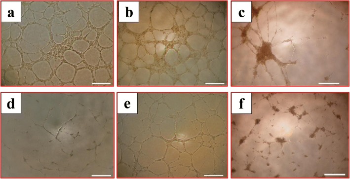
Fig. 8.
HUVEC tubule formation in presence of glucose and pMSCs. After 14 h, glucose-untreated pMSCs (a) and HUVECs cultured with 25% CMpMSC (b) able to form tube networks. HUVECs cultured with pMSCs alone did not form extensive tubule networks (c), and with 100 mM glucose alone (d) were unable to form tube networks. HUVECs cultured with 100 mM glucose and 25% CMpMSC (e) able to form tube networks, while culturing HUVECs with 100 mM glucose and pMSCs (f) failed to form extensive tube networks. Each experiment performed in triplicate using HUVECs (passage 3–5) and pMSCs (passage 2) from five independent umbilical cord tissues and placentae, respectively

