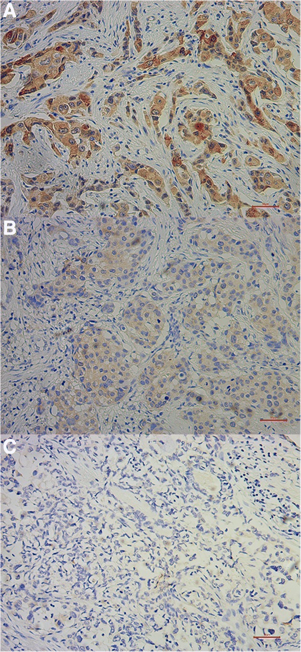
Fig. 1.

Immunohistochemical staining of MMP-9 in post-NAC samples of breast cancer. MMP-9 protein was detected with immunohistochemistry (IHC) using paraffin-embedded breast cancer samples collected after neoadjuvant chemotherapy. a Representative IHC images of strongly positive MMP-9 staining (200X). b Representative IHC images of moderate MMP-9 staining. c Representative IHC images of negative MMP-9 staining (200X). Scale bar: 50 μm
