Fig. 5.
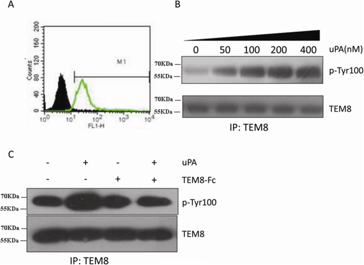
Stimulation of TEM8 phosphorylation by LMW-uPA. a HepG2 cells were incubated with (green, empty) or without (black, solid) an anti-TEM8 antibody, followed by detection with a FITC-labeled anti-IgG secondary antibody, measured by flow cytometry. b HepG2 hepatoma cells were serum-starved overnight and treated with LMW-uPA at the indicated doses. Cell lysates were immunoprecipitated with an anti-TEM8 antibody, followed by anti-p-Tyr100 immunoblotting. c HepG2 hepatoma cells were serum-starved overnight, pretreated with TEM8-Fc, and then stimulated with LMW-uPA. Cell lysates were immunoprecipitated with an anti-TEM8 antibody, followed by anti-p-Tyr100 immunoblotting

