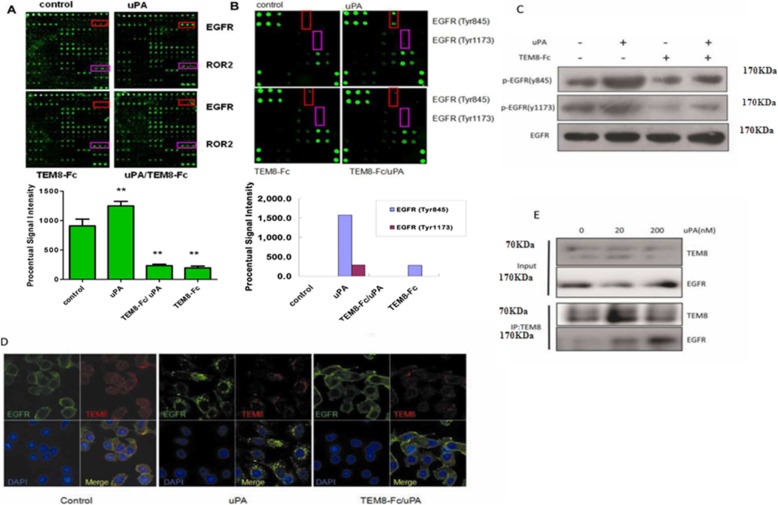
Fig. 6.
Induction of EGFR phosphorylation by uPA. a Treatment of HepG2 cells with uPA stimulates the phosphorylation of EGFR, which is blocked by TEM8-Fc, as detected by RayBio® Human RTK Phosphorylation Antibody Array 1. The fluorescent image (upper panel) and the quantification of that image (lower panel) are shown. b Treatment of HepG2 cells with uPA stimulates the phosphorylation of EGFR at Tyr845 and Tyr1173 as detected by RayBio® Human EGFR Phosphorylation Antibody Array 1. The fluorescent image (upper panel) and the quantification of that image (lower panel) are shown. c HepG2 cells were treated with uPA and/or TEM8-Fc as described in Materials and Methods, and then the cell lysates were subjected to immunoblotting with the indicated antibodies. d HepG2 cells were treated with uPA and/or TEM8-Fc, and then dual-color immunofluorescence was performed to determine the cellular localization of TEM8 and EGFR. e HepG2 cells were treated with uPA. The cell lysates were immunoprecipitated by TEM8 antibody and then subjected to immunoblotting with EGFR antibody. TEM8 proteins were knockdown in HepG2 cells and the cell lysates were then immunoblotted with the indicated antibodies

