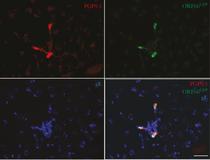
Figure 1.
VZVORF66.GFP infection of isolated enteric neurons. The longitudinal muscle and adherent myenteric plexus were dissected from the guinea pig small intestine and dissociated with collagenase. The ganglia were manually selected under microscopic control, isolated, and grown in culture. Peripheral blood mononuclear cells (PBMCs) were obtained from a donor guinea pig and cocultured with VZVORF66.GFP to infect T cells in the population. The VZVORF66.GFP-infected PBMCs were then cocultured with the infected neurons. After 1 week in culture, the cells were fixed and immunocytochemically examined as whole mounts. The immunoreactivities of varicella-zoster virus (VZV) glycoprotein E ([gE] blue), a neuronal marker, PGP9.5 (red), and green fluorescent protein (GFP) were visualized. Coincident immunostaining of PGP9.5 and GFP is found in neurons, which do not contain gE immunoreactivity. Many gE-immunoreactive nonneuronal cells are seen, many of which cluster around the neurons. The bar = 100 µm.

