Figure 7.

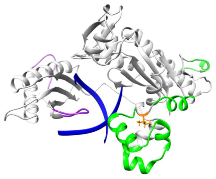
RMSF difference between WT and I236M Pol ι. Regions that show an increase(decrease) in RMSF in I236M Pol ι with respect to WT are shown in green(purple). The DNA substrate is shown in blue ribbons and the mutation site in orange sticks.

RMSF difference between WT and I236M Pol ι. Regions that show an increase(decrease) in RMSF in I236M Pol ι with respect to WT are shown in green(purple). The DNA substrate is shown in blue ribbons and the mutation site in orange sticks.