
Figure 2.
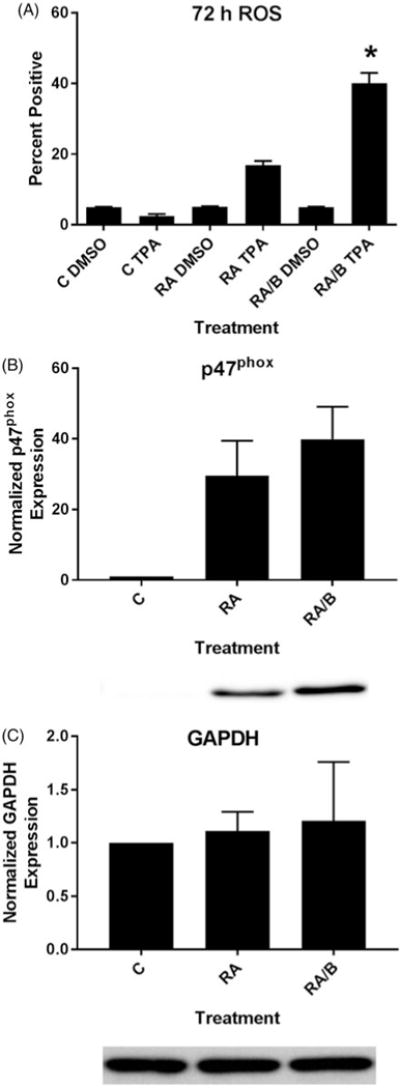
HL-60 cells treated with RA/bosutinib displayed enhanced respiratory burst and p47phox expression. (A) HL-60 cells were cultured in the presence of 1 μM RA or 1 μM RA and 0.25 μM bosutinib as indicated. Respiratory burst was analyzed by measuring inducible reactive oxygen species (ROS) production by flow cytometry using the 2′,7′-dichlorofluorescein (DCF) assay. Gates to determine percent increase of expression with treatment were set to exclude 95% of the DMSO-treated control population for each culture condition; TPA-treated samples show induced ROS (n = 3). Error bars indicate SEM. *p < .05 comparing RA-treated samples to RA/bosutinib-treated samples. Two-tailed paired-sample t-tests were used to determine significance. (B) HL-60 cells were cultured for 48 h in the presence of 1 μM RA or 1 μM RA and 0.25 μM B as indicated and whole cell lysate was collected. Twenty five microgram of lysate per lane was run. Western blots of PAGE-resolved lysates were probed for p47phox (n = 3). Films were scanned and bands of interest were quantified using NIH ImageJ. Error bars indicate SEM. A representative blot, cropped to show only the band of interest, is included. (C) Western blots of GAPDH were used as loading controls following the procedure described above.