
Figure 3.
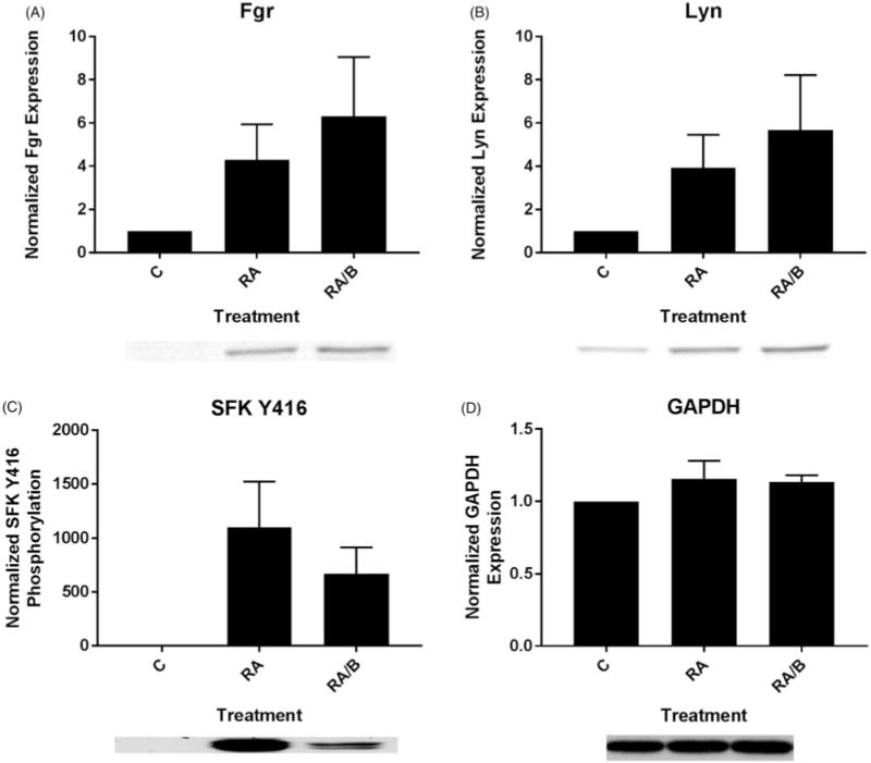
Bosutinib enhances RA-induced SFK expression and diminishes SFK phosphorylation. (A) HL-60 cells were cultured for 48 h in the presence of 1 μM RA or 1 μM RA and 0.25 μM bosutinib (B) as indicated and whole cell lysate was collected. Twenty five microgram of lysate per lane was run. Western blots of PAGE-resolved lysates were probed for Fgr (n = 3). Films were scanned and bands of interest were quantified using NIH ImageJ. Error bars indicate SEM. A representative blot, cropped to show only the band of interest, is included. (B) Western blots of Lyn following the procedure described above. (C) Western blots of phosphorylated pan-Y416 SFK following the procedure described above. (D) Western blots of GAPDH were used as loading controls following the procedure described above.