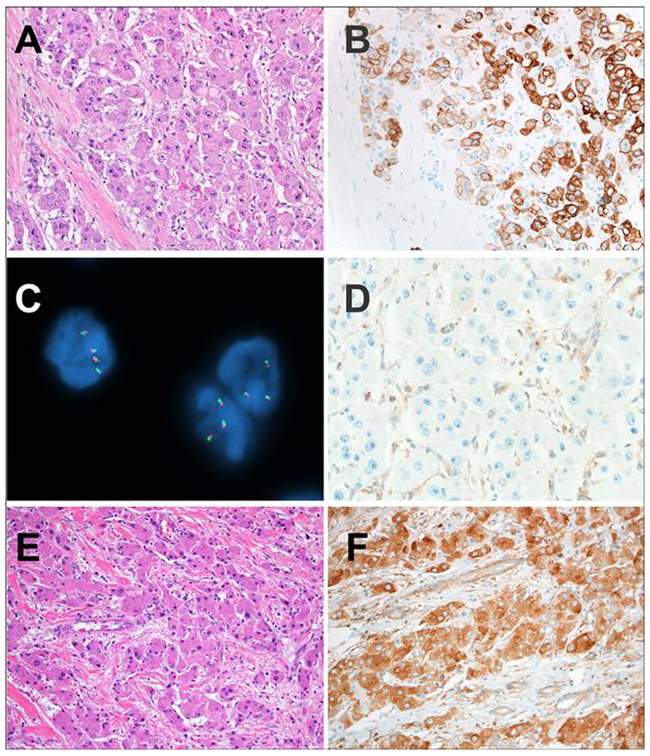
Figure 1 –
A: The liver tumors in the patients with the Carney complex were fibrolamellar carcinomas. The classic histologic features of fibrolamellar carcinoma are shown in this case including eosinophilic cells with abundant cytoplasm and prominent macronucleoli. Pale bodies were seen. Lamellar bands of fibrosis traversed the tumor.
B: The tumor cells of fibrolamellar carcinoma in patients with the Carney complex were positive for Keratin 7, as expected for fibrolamellar carcinoma
C: PRKACA break apart FISH revealed intact PRKACA loci with closely opposed red and green signals in Carney complex-associated fibrolamellar carcinomas. This finding indicates absence of PRKACA rearrangement in the tumor cells.
D: PRKAR1A expression was absent in the tumor cells of fibrolamellar carcinomas in patients with the Carney complex. The Kupffer cells served as an internal control and revealed retained expression.
E: Sporadic control fibrolamellar carcinomas also showed the classic morphology of fibrolamellar carcinoma.
F: PRKAR1A expression was retained in the neoplastic cells of all sporadic control fibrolamellar carcinomas.

