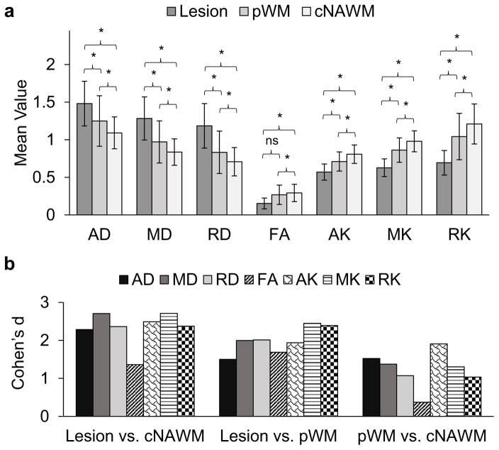
Figure 3.
a) Average values for mean, axial and radial diffusivity (MD, AD, and RD) and kurtosis (MK, AK, and RK) and fractional anisotropy (FA) in the solid lesion, surrounding perilesional white matter (pWM) and contralateral normal appearing white matter (cNAWM) in 19 patients with low-grade gliomas. Mean, axial and radial diffusivity have units 10−3 mm2/sec; mean, axial and radial kurtosis and fractional anisotropy are dimensionless. The significance of paired differences are shown by * denoting False Discovery Rate (FDR) corrected p <0.001. b) Effect size calculated using Cohen’s d for paired differences.

