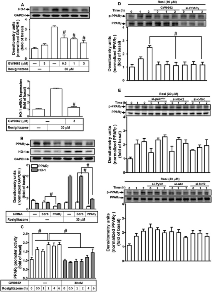
Figure 9.

Rosiglitazone induces HO‐1 expression through a PPARγ‐dependent manner. (A) HPAEpiCs were pretreated with various concentrations of GW9662 for 1 h and then incubated with vehicle or rosiglitazone (30 μM) for 16 h. The levels of HO‐1 and GADPH proteins were determined by Western blot. (B) Cells were transfected with PPARγ siRNA and then incubated with rosiglitazone for 16 h. The levels of PPARγ, HO‐1 and GAPDH were detected by Western blot. (C) PPARγ‐RE reporter construct‐transfected cells were pretreated with GW9662 (30 nM) for 1 h and then incubated rosiglitazone (30 μM) for 2 h. PPARγ RE promoter luciferase activity was determined in the cell lysates. (D, E) HPAEpiCs were pretreated with GW9662 and transfected with PPARγ, p47phox, NOX2, c‐Src, Pyk2, Akt or Nrf2 siRNA, and then incubated with vehicle or rosiglitazone (30 μM) for the indicated time intervals. Western blot was used to detect the phosphorylation of PPARγ and the levels of total PPARγ. Data are expressed as mean ± SEM, from five independent experiments (n = 5). # P < 0.05, significantly different from rosiglitazone alone (A, D); or significantly different as indicated (B–D).
