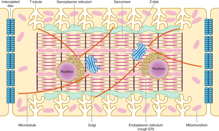
FIGURE 1.
Schematic illustration of the internal structures of an adult ventricular cardiomyocyte. T-tubules, which are enriched with voltage-gated L-type calcium channels, are positioned closely near the sarcoplasmic reticulum, the primary internal calcium store. Sarcomeres form myofibrils, which are responsible for cardiomyocyte contraction upon calcium release. The Golgi apparatus and microtubules serve as the “loading dock” and “highways,” respectively, to deliver ion channels to specific subdomains on the plasma membrane. Mitochondria provide the energy needed for the contraction of cardiomyocytes. Intercalated discs located at the longitudinal sides of each ventricular cardiomyocyte mediate the cell-to-cell propagation of action potentials.

