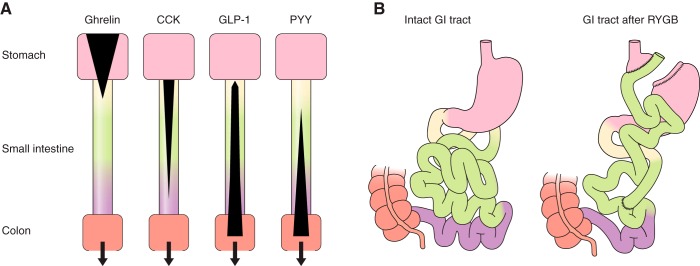
FIGURE 1.
Schematic depictions of the localization of enteroendocrine cells and changes after RYGB. A: distribution of enteroendocrine cells secreting ghrelin, CCK, GLP-1, and PYY in the stomach (pink), duodenum (yellow), jejunum (green), and ileum (violet). Black areas indicate the relative densities of expression of enteroendocrine cells producing the hormones indicated. Enteroendocrine cells secreting particular hormones were initially categorized histologically, e.g., I cells for CCK, L cells for enteroglucagons and PYY, etc. (166, 567, 591). It is now clear, however, that this categorization is not a reliable guide to hormone secretion. Rather, individual enteroendocrine cells secrete variable mixtures of hormones (231, 303, 597, 738). Bottom salmon rectangle, proximal large intestine. B: intact gastrointestinal tract (left) and gastrointestinal rearrangement after RYGB (right). Pink areas are stomach, salmon areas are large intestine (∼1.5 m long in healthy adults), yellow is duodenum (typically ∼25 cm long), green is jejunum (∼2–3 m), and violet is ileum (∼3–4 m). For RYGB, the stomach is divided into a small upper pouch with a volume of ∼25 ml and an isolated gastric remnant, the small intestine is divided ∼50 cm from the pylorus, and the distal limb of the small intestine (Roux or alimentary limb) is brought up to the gastric pouch and connected to it by an end-to-side gastroenterostomy. As a result, ingested food enters the small gastric pouch and empties directly into the jejunum. The gastric remnant and isolated ∼50 cm of small intestine (“biliopancreatic limb”) is connected to the jejunum ∼150 cm distal to the gastroenterostomy. The small intestine distal to the anastomosis is called the common channel.

