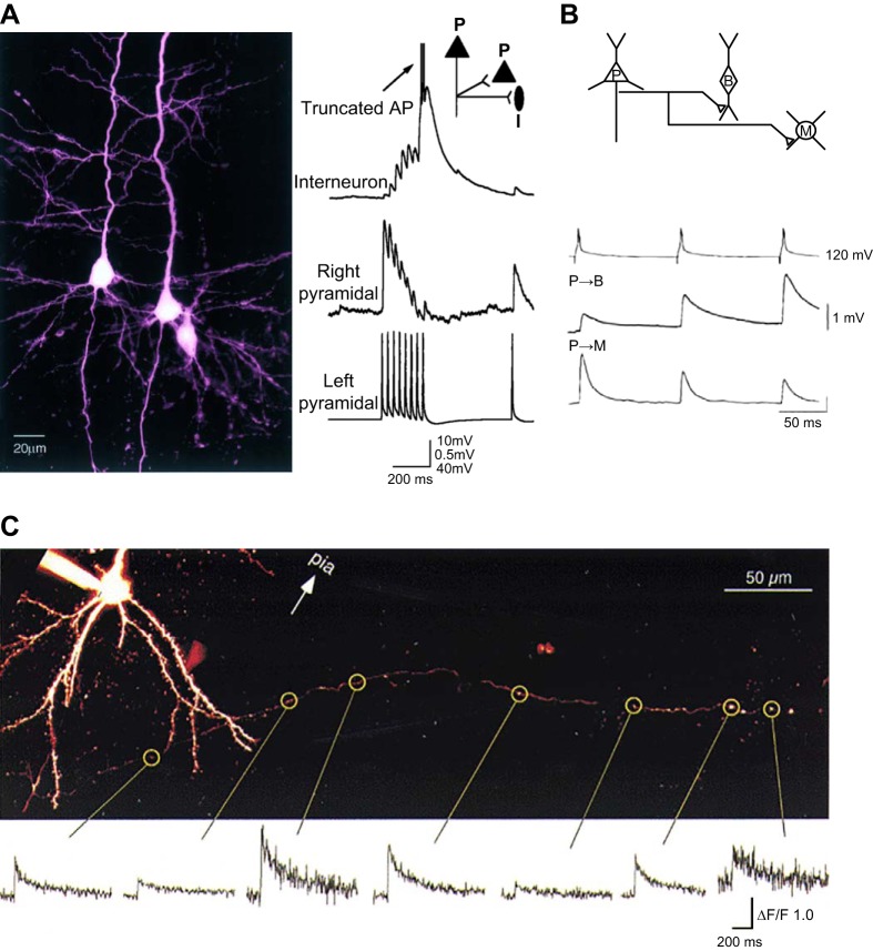
FIGURE 16.
Target-cell-dependent inhibitory transmission. A, left: image of three biocytin-filled neurons in layer 5 somatosensory cortex. The pyramidal neuron on the left innervated another pyramidal neuron and a bipolar interneuron, both on the right. A, right: single-trial responses (30 Hz) to the same action potential train (evoked in the presynaptic left pyramidal cell) for the simultaneously recorded postsynaptic interneuron and pyramidal cell targets. Note the strong frequency facilitation and depression of synaptic events in the interneuron and pyramidal postsynaptic targets, respectively. B: data from a triple-patch recording in layer2/3 somatosensory cortex, revealing differential short-term plasticity in two classes of interneurons innervated by a single pyramidal neuron. Three action potentials evoked at 10 Hz in the presynaptic pyramidal cell (top trace), evoked short-term facilitation of unitary EPSPs evoked in the bitufted cell (middle trace, P-B connection), whereas the amplitude of EPSPs evoked simultaneously in the multipolar cell decreased (bottom trace, P-M connection). C: presynaptic Ca2+ transients at divergent release sites of the same axon exhibit target-cell dependence. A single layer 2/3 pyramidal cell loaded with Ca2+ indicator (upper fluorescence image) displays a large degree of heterogeneity in single action potential-evoked Ca2+ transients (bottom traces) at various boutons (circles) along a single axon collateral. [A from Markram et al. (749). B from Reyes et al. (940) with permission from Nature Neuroscience. C from Koester and Sakmann (591) with permission from Journal of Physiology.]

