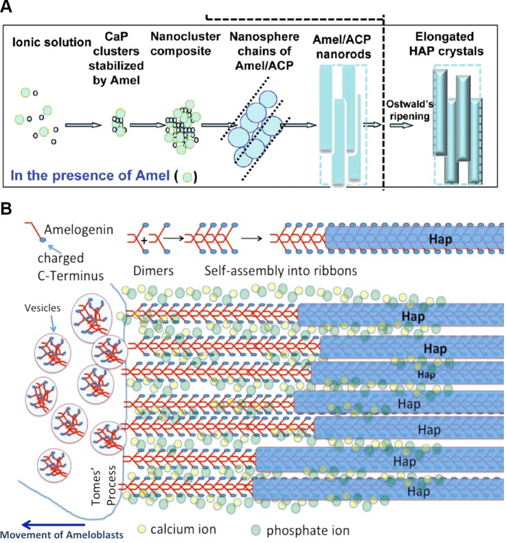
FIGURE 9.
Proposed models of amelogenin-directed growth of apatite mineral during secretory stage of amelogenesis. Comparison of two models of amelogenin-directed growth of apatite mineral during secretory stage of amelogenesis. A: nanosphere model. Amelogenin protein is secreted into the extracellular space and assembles into nanospheres. Amelogenin stabilizes prenucelation clusters of calcium phosphates. Nanospheres align into chainlike structures along which amorphous calcium phosphate develops and through a ripening process transforms into crystalline Hap. Nanospheres act as spacers, as originally proposed by Fincham et al. (150). [A from Yang et al. (662), copyright 2010 American Chemical Society.] B: nanoribbon-directed crystal growth. Amelogenin is secreted from vesicles, possibly in the form of antiparallel dimers. Nanoribbon assembly is triggered by formation of ion bridges across dimers with Ca2+ and PO43−. Dimers are added to the existing amelogenin ribbons as soon as they are exocytosed and thus elongate the ribbons as the ameloblasts migrate away from the mineralization front. Hap mineral forms at a narrow distance from the cell membrane in form of thin ribbons that develop on the backbone of the protein ribbons. Full-length amelogenin does not induce mineral formation directly, and the mechanism of mineral nucleation is not known. Interaction with other non-amelogenin molecules and/or the processing by MMP20 may be required for guided growth of apatite onto amelogenin ribbons.

