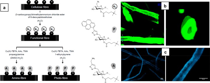
Figure 2.
(a) Synthesis scheme for the preparation of cellulose fibers decorated with photoactive molecules (photofibers) and amino functional groups (amino fibers) prepared from cellulose fibers decorated with azide functions (reactive fibers). Cu(II)-TBTA, Cu(II)-tris[(1-benzyl-1H-1,2,3-triazol-4-yl)methyl]amine complex solution; AAc, ascorbic acid; TMA, triethylammonium acetate buffer. (b) Section (xy plane) of scanned xyz volume of photofibers obtained with TPM (left). A 3D rendering of a cross-section of an individual fiber at the position marked as “ROI2” on the left figure (right). The figures illustrate the dense labeling of the fibers and the preserved 3D shape during the activation and labeling. (c) Image of a photofiber observed with an Olympus BX60 epi-fluorescence microscope at 10× (left) and 40× magnifications (right). Excitation filter, 330–385 nm; dichroic mirror, 400 nm; and barrier filter, >420 nm. Reprinted from ref (19) with permission from Elsevier.

