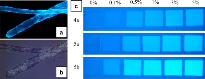
Figure 8.

Visualization of fluorescent pulp fibers by an epifluorescence microscope under UV light exposure (a) and white light (b). The fibers were modified with 5b, and the dosage was 2% (w/w). N-(4-Butanoic acid)-1,8-naphthalimide-(3-carboxypropyl)trimethylammonium chloride ester of cellulose (5b, DSphoto 0.22, DScat 0.33). (c) Picture of fiber hand-sheets under black light illumination. The quadrates and the background are made of treated FMCDs and reference fibers, respectively. N-(3-Propanoic acid)-1,8-naphthalimide-(3-carboxypropyl)trimethylammonium chloride ester of cellulose (4a, DSphoto 0.07, DScat 0.31) and N-(4-butanoic acid)-1,8-naphthalimide-(3-carboxypropyl)trimethylammonium chloride esters of cellulose (5a, DSphoto 0.11, DScat0.32; 5b, DSphoto 0.22, DScat 0.33). Reprinted from ref (46) with permission from the American Chemical Society.
