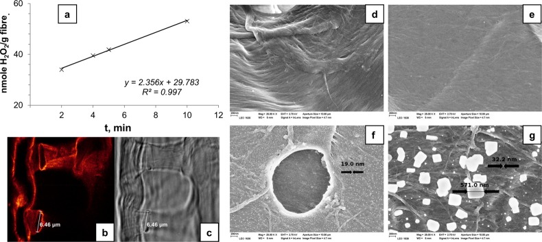
Figure 9.
(a) Enzyme activity shown by biocatalytic 673 fibers FG*. STED image of a biocatalytic fiber cross-section bearing free enzymes labeled with Abberior STAR 635 (b) and the corresponding transmission image (c). Dye/enzyme ratio of 1.4. Picture dimensions: 32.53 μm × 32.53 μm (b, c). Field emission scanning electron microscope (FESEM) pictures of the reference fibers (d, e) and the fibers obtained after chemical reaction between the functional fibers (F), the immobilized enzymes (G*), and GA (f, g). All images are at 25K magnification. Reprinted from ref (50) with permission from the American Chemical Society.

