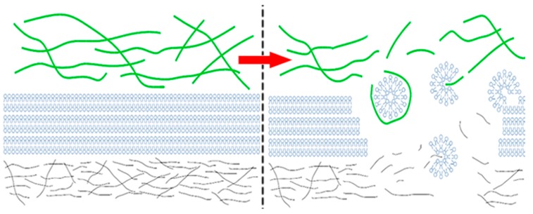
Figure 1.
Graphical simplification of a synovial joint organ system. The left-hand side depicts normal synovial fluid conditions with longer hyaluronic acid (HA) chains and mature HA networks (green), phospholipid (PL) bilayers of the oligolamellar surface-active phospholipid layer (blue), and the superficial zone of articular cartilage (grey). The right-hand side depicts osteoarthritic synovial fluid conditions with correspondingly shorter HA chains, rudimentary HA networks, PL micelles, and a damaged superficial zone upon which the surface-active phospholipid layer cannot form.

