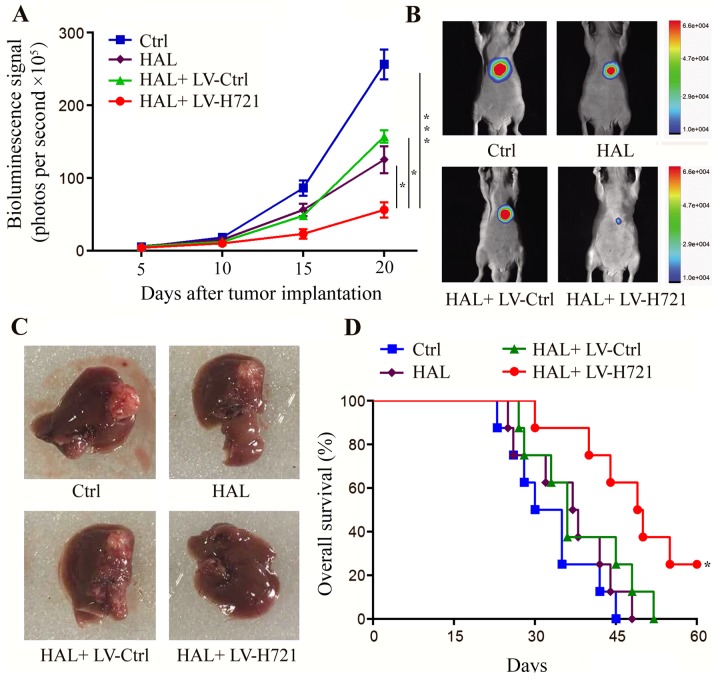
Figure 5.
Effect of hypoxia inducible factor-1α disruption on tumor growth and progression in the mouse orthotopic model. At 5 days after tumor cell implantation, the animals were randomly divided into four groups. (A) Longitudinal monitoring was plotted by the detection of the mean bioluminescence signals on days 5, 10, 15 and 20 after cell implantation (error bars represent the standard error of the mean). (B) Live imaging of the representative tumors on day 20. (C) Tumors of mice sacrificed on day 20, indicating that the tumor volume was evidently decreased in the HAL + LV-H721 group. (D) Animal median survival in each group plotted by the log-rank test. Statistically significant differences were denoted between HAL + LV-H721 group and the other three groups correspondingly (Ctrl, HAL, and HAL + LV-Ctrl groups, all *P<0.05). Data are representative of four independent experiments. *P<0.05 and ***P<0.001. HAL, hepatic artery ligation; Ctrl, control; LV, lentivirus.

