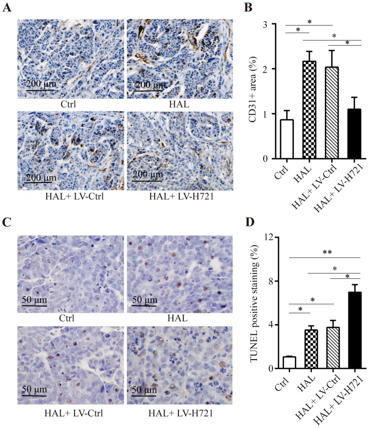
Figure 6.
Immunohistochemical analysis of microvascular density and TUNEL assay in the mouse orthotopic tumor model. Tumor tissues were acquired at 20 days after the different treatments. (A) Monoclonal anti-CD31 antibody staining of the section to examine the microvascular density with ×20 magnification, and (B) CD31-positive cells. Five sections were counted for each slide. (C) TUNEL assay was performed on the SMMC-7721-Fluc-induced orthotopic hepatocellular carcinoma model in mice with ×20 magnification, and (D) the percentage of TUNEL-positive cells is presented. Values represent the mean ± standard error of the mean, and are representative of four independent experiments. *P<0.05, **P<0.01. HAL, hepatic artery ligation; Ctrl, control; LV, lentivirus.

