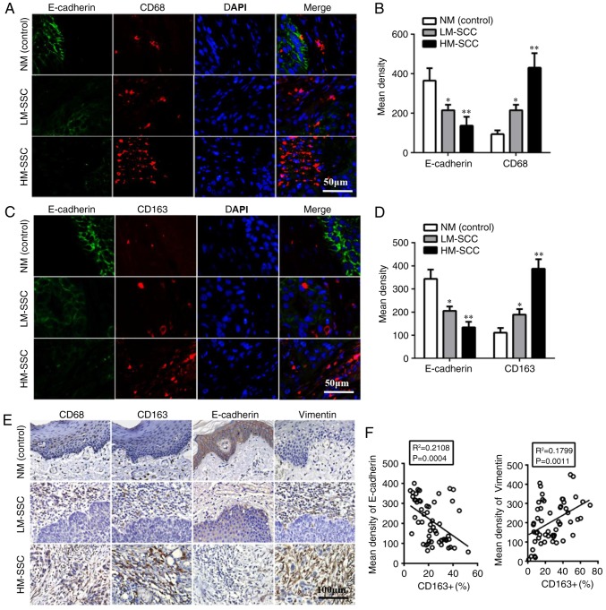
Figure 1.
Tumor associated macrophages biomarkers are associated with EMT-associated proteins in HNSCC. (A) Detection of CD68 and E-cadherin expression by double immunofluorescent staining in HNSCC tissues. Magnification, ×200. (B) Quantitative data of CD68 and E-cadherin expression from immunofluorescent staining in HNSCC tissues. (C) Detection of CD163 and E-cadherin by double immunofluorescent staining in HNSCC tissues. Magnification, ×200. (D) Quantitative data of CD163 and E-cadherin from immunofluorescent staining in HNSCC tissues. Data are expressed as the mean ± standard error of the mean. *P<0.05 and **P<0.01 vs. NM (control). (E) Detection of CD68, CD163 and EMT-associated protein expression by immunohistochemical staining in HNSCC tissues. Magnification, ×200. (F) Pearson correlation analysis of EMT-associated protein and CD163+ expression. HNSCC, head and neck squamous cell carcinoma; EMT, epithelial to mesenchymal transition; CD, cluster of differentiation; HM-SCC, HNSCC with high macrophages; LM-SCC, HNSCC with low macrophages; NM, normal mucosa.

