Figure 6.
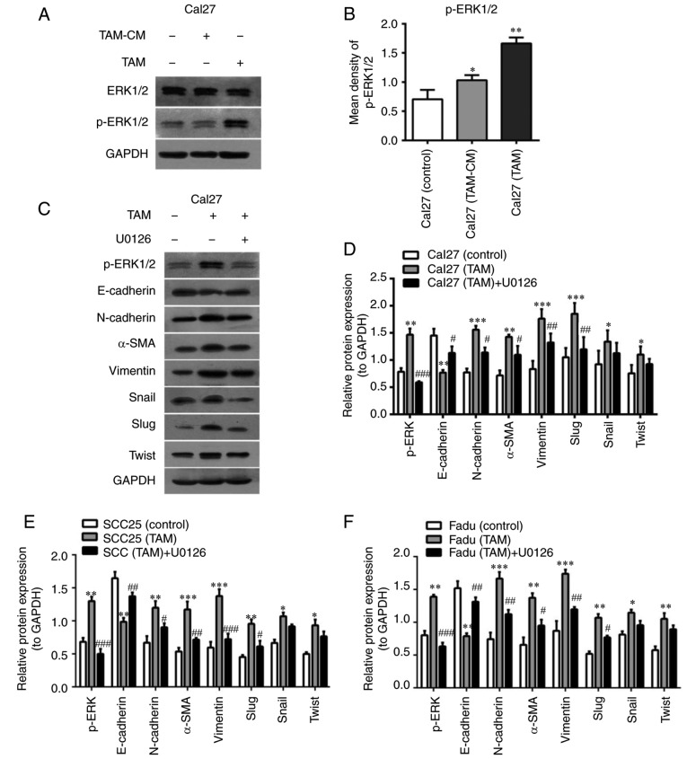
TAMs induce EMT in HNSCC cells via the ERK1/2 signaling pathway. (A) The ERK1/2 signaling pathway was detected in Cal27 cells in the direct and indirect co-culture system by western blot analysis. (B) Expression of the ERK1/2 signaling pathway was quantified. (C) A specific inhibitor of ERK1/2 (U0126; 10 µg/ml) was added to the co-culture system of TAMs and Cal27 cells for 24 h. Cal27 cells were labeled with carboxyfluorescein diacetate succinimidyl ester and sorted using fluorescence-activated cell sorting. ERK1/2 pathway activating and EMT-associated proteins were measured in Cal27 cells by western blotting. (D-F) Quantitative analysis of p-ERK1/2 and EMT-associated proteins in (D) Cal27, (E) SCC25 and (F) Fadu cells. Data are expressed as the mean ± standard error of the mean. *P<0.05, **P<0.01 and ***P<0.001 vs. Control; #P<0.05, ##P<0.01 and ###P<0.001 vs. tumor cells co-cultured with TAMs. TAMs, tumor associated macrophages; EMT, epithelial to mesenchymal transition; ERK1/2, extracellular signal-regulated protein kinase 1/2; p-, phosphorylated; α-SMA, α-smooth muscle actin; CM, conditional media.

