Figure 2.

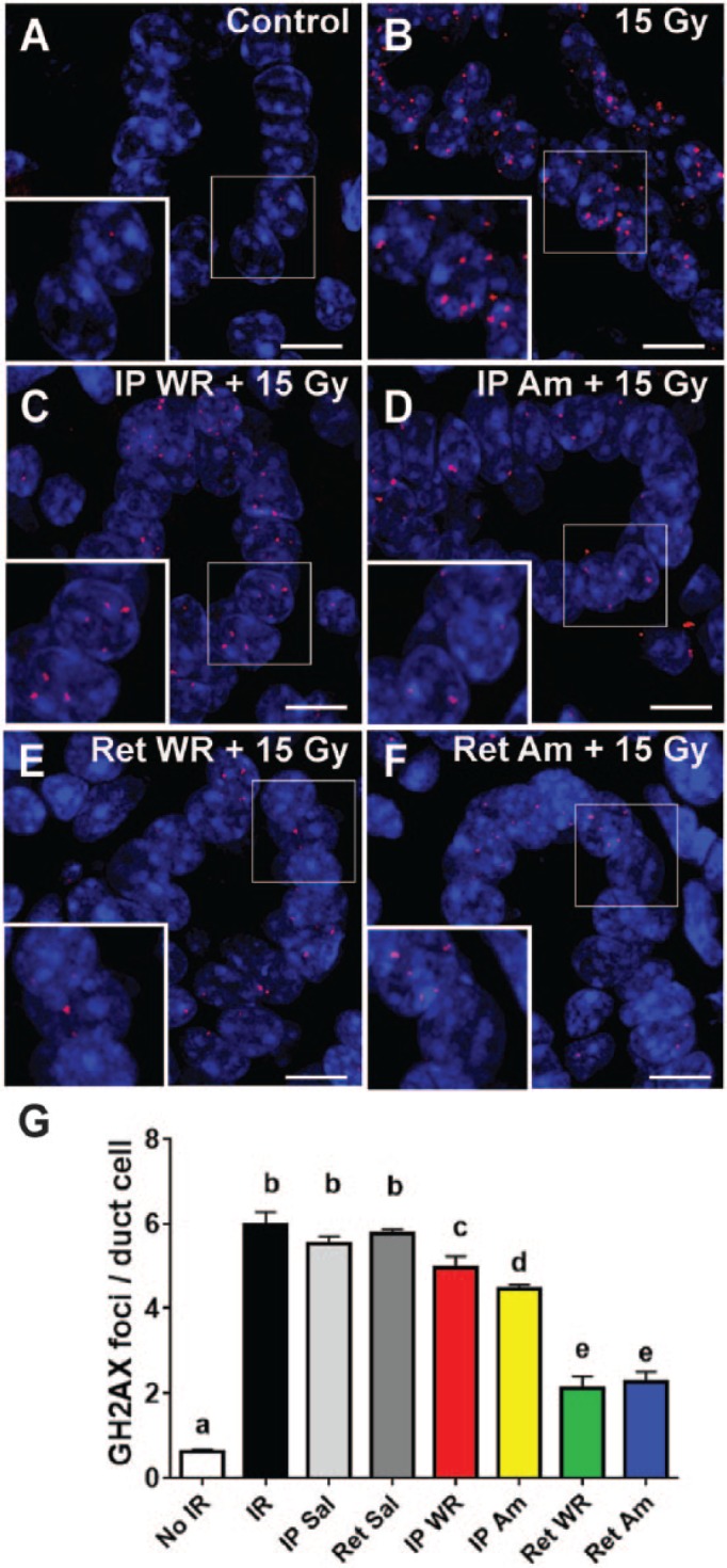
Immunofluorescent staining for γH2AX (red punctate dots) localizes to sites of DNA damage in nuclei of duct cells at 48 h after 15 Gy irradiation (scale bars = 5 μm). Sections of submandibular gland isolated from control (A), 15 Gy (B), IP WR+ 15 Gy (C), IP Am + 15 Gy (D), Ret WR + 15 Gy (E), and Ret Am + 15 Gy (F) labeled with antibody to γH2AX. Nuclei stained with DAPI. (G) Quantification of γH2AX foci, per duct cell per field. All groups are significantly different from group a. Group e is significantly different from all other groups. Group c is not different from all members of group b. Group d is significantly different from all groups except c (mean ± SEM, n = 5, 1-way analysis of variance with Tukey’s post hoc test for multiple comparisons). Am, amifostine; IP, intraperitoneal; IR, irradiation; Ret, retroductal; Sal, saline; WR, WR-1065.
