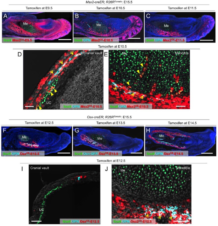
Figure 2.
Msx2-creER+ cells become diverse types of mesenchymal cells in fetal development. (A) Sagittal sections of Msx2-creER; R26RTomato mandibles at E15.5 (tamoxifen at E9.5) stained for Sox9 and nuclei. Me, Meckel’s cartilage. Green: Sox9-Alexa488; red: tdTomato; blue: 4′,6-diamidino-2-phenylindole (DAPI); gray: differential interference contrast (DIC). Scale bars: 400 µm. (B) Sagittal sections of Msx2-creER; R26RTomato mandibles at E15.5 (tamoxifen at E10.5) stained for Sox9, osteopontin (OPN), and nuclei. Me, Meckel’s cartilage. Light blue: OPN–Alexa 647; green: Sox9–Alexa 488; red: tdTomato; blue: DAPI; gray: DIC. Scale bars: 400 µm. (C) Sagittal sections of Msx2-creER; R26RTomato mandibles at E15.5 (tamoxifen at E11.5) stained for Sox9, OPN, and nuclei. Me, Meckel’s cartilage. Light blue: OPN–Alexa 647; green: Sox9–Alexa 488; red: tdTomato; blue: DAPI; gray: DIC. Scale bars: 400 µm. (D) Sagittal sections of Msx2-creER; R26RTomato cranial vault at E15.5 (tamoxifen at E10.5) stained for Sox9, OPN, and nuclei. Arrows: OPN+tdTomato+ osteoblasts; arrowheads: Sox9+tdTomato+ chondrocytes. UC, underlying cartilage. Light blue: OPN–Alexa 647; green: Sox9–Alexa 488; red: tdTomato; gray: DAPI and DIC. Scale bars: 50 µm. (E) Sagittal sections of Msx2-creER; R26RTomato mandible at E15.5 (tamoxifen at E10.5) stained for Sox9, OPN, and nuclei. Magnified view of the dotted area in (B). Me, Meckel’s cartilage. Arrows: OPN+tdTomato+ osteoblasts; arrowheads: Sox9+tdTomato+ chondrocytes and perichondrial cells. Light blue: OPN–Alexa 647; green: Sox9–Alexa 488; red: tdTomato; gray: DAPI and DIC. Scale bars: 50 µm. (F) Sagittal sections of Osx-creER; R26RTomato mandibles at E15.5 (tamoxifen at E12.5) stained for Sox9, OPN, and nuclei. Arrows: OPN+tdTomato+ osteoblasts. Me, Meckel’s cartilage. Light blue: OPN–Alexa 647; green: Sox9–Alexa 488; red: tdTomato; blue: DAPI; gray: DIC. Scale bars: 400 µm. (G) Sagittal sections of Ocn-GFP; Osx-creER; R26RTomato mandibles at E15.5 (tamoxifen at E13.5) stained for Sox9 and nuclei. Me, Meckel’s cartilage. Light blue: Ocn-GFP; green: Sox9–Alexa 647; red: tdTomato; blue: DAPI; gray: DIC. Scale bars: 400 µm. (H) Sagittal sections of Osx-creER; R26RTomato mandibles at E15.5 (tamoxifen at E14.5) stained for Sox9, OPN, and nuclei. Me, Meckel’s cartilage. Light blue: OPN–Alexa 647; green: Sox9–Alexa 647; red: tdTomato; blue: DAPI; gray: DIC. Scale bars: 400 µm. (I) Sagittal sections of Osx-creER; R26RTomato cranial vault at E15.5 (tamoxifen at E12.5) stained for Sox9, OPN, and nuclei. UC, underlying cartilage. Light blue: OPN–Alexa 647; green: Sox9–Alexa 488; red: tdTomato; gray: DAPI and DIC. Scale bars: 50 µm. (J) Sagittal sections of Osx-creER; R26RTomato mandibles at E15.5 (tamoxifen at E12.5) stained for Sox9, OPN, and nuclei. Magnified view of the dotted area in (F). Me, Meckel’s cartilage. Arrows: OPN+tdTomato+ osteoblasts. Light blue: OPN–Alexa 647; green: Sox9–Alexa 488; red: tdTomato; gray: DAPI and DIC. Scale bars: 50 µm.

