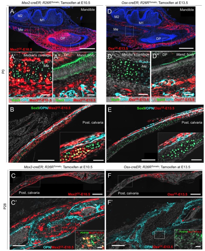
Figure 3.
Msx2-creER+ early mesenchymal cells exhibit broader differentiation potential than Osx-creER+ osteoblast precursors. (A–A′′) Sagittal sections of Msx2-creER; R26RTomato mandibles at P0 (tamoxifen at E10.5) stained for Sox9, osteopontin (OPN), and nuclei. A′: Meckel’s cartilage; A′′: mandibular body. DP, dental pulp; M1, mandibular first molar; M2, mandibular second molar; Me, Meckel’s cartilage. Light blue: OPN–Alexa 647; green: Sox9–Alexa 488; red: tdTomato; gray: 4′,6-diamidino-2-phenylindole (DAPI) and differential interference contrast (DIC). Scale bars: 500 µm (A) and 50 µm (A′–A′′). (B) Sagittal sections of Msx2-creER; R26RTomato posterior cranial vault at P0 (tamoxifen at E10.5) stained for Sox9, OPN, and nuclei. Inset: magnified view of the transitional area. UC, underlying cartilage. Light blue: OPN–Alexa 647; green: Sox9–Alexa 488; red: tdTomato; gray: DAPI and DIC. Scale bars: 200 µm and 50 µm (inset). (C–C′) Sagittal sections of Msx2-creER; R26RTomato posterior cranial vault at P28 (tamoxifen at E10.5) stained for OPN, periostin (POSTN), and nuclei. Inset: magnified view of the dotted area of the suture. Light blue: OPN–Alexa 647; green: POSTN–Alexa 488; red: tdTomato; gray: DAPI and DIC. Scale bars: 500 µm and 20 µm (inset). (D–D′′) Sagittal sections of Osx-creER; R26RTomato mandibles at P0 (tamoxifen at E13.5) stained for Sox9, OPN, and nuclei. DP, dental pulp; M1, mandibular first molar; M2, mandibular second molar; Me, Meckel’s cartilage. Light blue: OPN–Alexa 647; green: Sox9–Alexa 488; red: tdTomato; gray: DAPI and DIC. Scale bars: 500 µm (D) and 50 µm (D′–D′′). (E) Sagittal sections of Osx-creER; R26RTomato posterior cranial vault at P0 (tamoxifen at E13.5) stained for Sox9, OPN, and nuclei. Inset: magnified view of the transitional area. UC, underlying cartilage. Light blue: OPN–Alexa 647; green: Sox9–Alexa 488; red: tdTomato; gray: DAPI and DIC. Scale bars: 200 µm and 50 µm (inset). (F–F′) Sagittal sections of Osx-creER; R26RTomato posterior cranial vault at P28 (tamoxifen at E13.5) stained for OPN, POSTN, and nuclei. Inset: magnified view of the dotted area of the suture. Light blue: OPN–Alexa 647; green: POSTN–Alexa 488; red: tdTomato; gray: DAPI and DIC. Scale bars: 500 µm and 20 µm (inset).

