
FIG. 2.
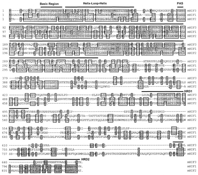
Alignment of three HIFs. The amino acid sequence of mouse HIF3α is aligned with that of mouse HIF1α and HIF2α using the CLUSTAL method. The positions of amino acids are shown on the left side. The basic region (BR), helix–loop–helix (HLH), PAS, and the hypoxia-responsive domains (HRDs) are marked with dark lines above the sequence. Conserved residues are boxed and in light gray.