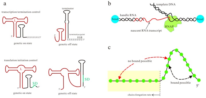
Figure 1.
The transcription and translation control mechanisms of riboswitches (a), where the aptamer sequence and Shine–Dalgarno (SD) sequence are colored brown and green respectively; L is the ligand. A schematic of the optical-trapping assay and the kinetic Monte Carlo simulation is shown in (b,c). In (c), nucleotides (green circles) out of the tube (yellow) are possible to form base pairs. The kinetic Monte Carlo method grows the RNA chain with a constant rate, allowing more and more base pairs to form or open in the available sequence.

