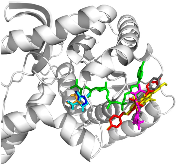
Figure 3.
The conformation of OXQ crystal structure (dark blue stick presentation), docked OXQ (light blue stick presentation), lactate (brown stick presentation) and test compound 3a, 3f, 4a and 4g (grey, red, pink and yellow stick presentation, respectively) in parasite lactase dehydrogenase (pLDH). Green stick is the NAD+ as the co-factor of pLDH.

