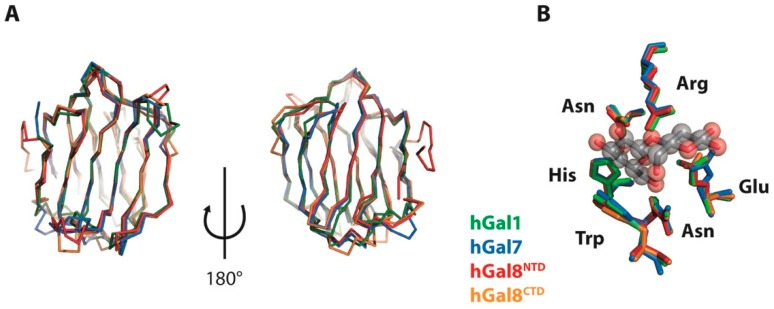
Figure 1.
Superimposition of X-ray crystal structures of hGal1, hGal7, hGal8NTD, and hGal8CTD. (A) Ribbon representation of the crystal structures of hGal1 (protein database entry (PDB ID): 1W6N; green), hGal7 (PDB ID: 1BKZ; blue), hGal8NTD (PDB ID: 3VKN; red), and hGal8CTD (PDB ID: 3OJB; orange) are shown here on two sides; (B) Detailed views of structural alignment of lactose binding residues. The side-chains of individual residues are coloured in the same way.

