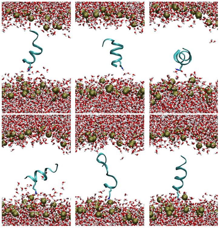
Figure 3.
Representative structures for each one of the three clusters that characterize the configurations sampled in the global minimum of the PMF profile, as obtained using the g_cluster tool in the GROMACS software package. The structures reported above refer to the TempL peptide, those reported below to Q3K-TempL. The phosphorus atoms of the lipid-A, marking the position of the lipid polar heads, are indicated as gold spheres. The peptide backbone is reported as a cyan ribbon, the water molecules and the third residue of the peptides (Q and K in TempL and Q3K-TempL, respectively) are reported as sticks, with O, N and H atoms colored in red, blue and white, respectively. For the sake of clarity, the lipid tails are not reported.

