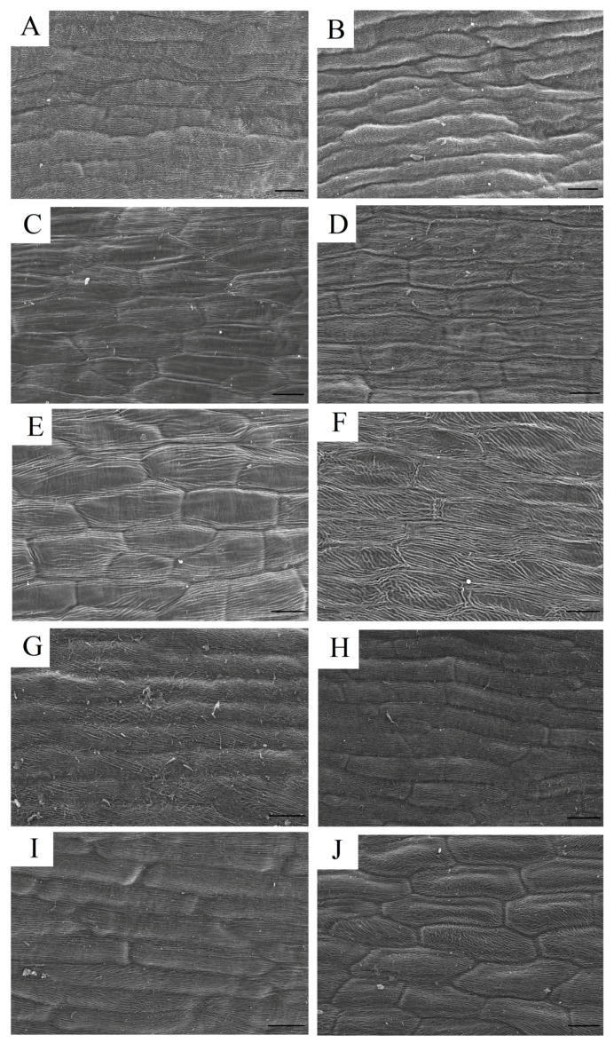
Figure 3.
Scanning electron micrograph (SEM) of papillate cells from the outer epidermis of petals from the materials. Bar = 40 μm. ((A,B), (E,F) and (G,H)) the upper and lower epidermis of the background petals of P. rockii, P. ostii and the F1 progeny, respectively. ((C,D) and (I,J)) the upper and lower epidermis of the variegated petals of P. rockii and the F1 progeny, respectively.

