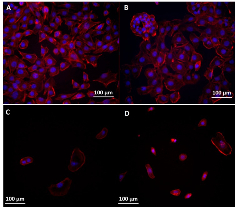
Figure 10.
Morphology of the cytoskeleton and nuclei in BT 474 cells: (A) control; (B) exposed to the highest concentration of Fe3O4; (C) exposed to the highest concentration of Fe3O4@GEM and (D) exposed to the highest concentration of GEM, at 24 h (0.09 mg/mL equivalent concentration of GEM). The cytoskeleton and nuclei were stained with red X-phalloidin and Hoechst.

