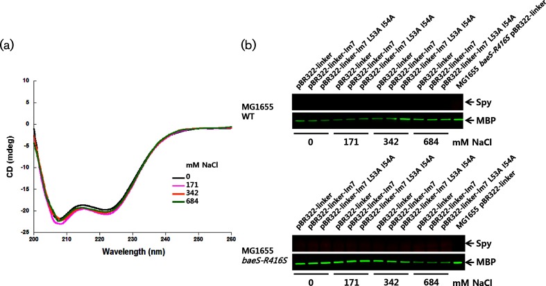
Fig. 2.
The secondary structure of Spy and its expression level was monitored. (a) The secondary structure of the Spy protein was monitored by CD spectroscopy. (b) Total cells were prepared from cells grown on LB agar media with different NaCl concentrations, separated by 4–12 % gradient SDS-PAGE. The Spy protein level was detected by Western blotting using Spy-specific antiserum. Maltose-binding protein (MBP) was also monitored as a reference protein using MBP polyclonal antibody (NEB). Spy and MBP are indicated by arrows.

