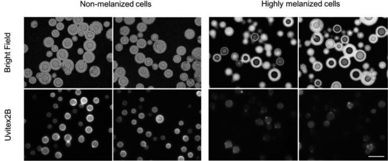
Fig. 1.
C. neoformans melanized cells show dimmed Uvitex fluorescent signal. Light microscopy of C. neoformans cells stained with India Ink (top panel) or with 5 µl of Uvitex (stock 20 mg ml−1) in the dark for 10 min at room temperature (bottom panel). Cell images were obtained using 100× oil-immersed objective. Scale bar, 10 µm.

