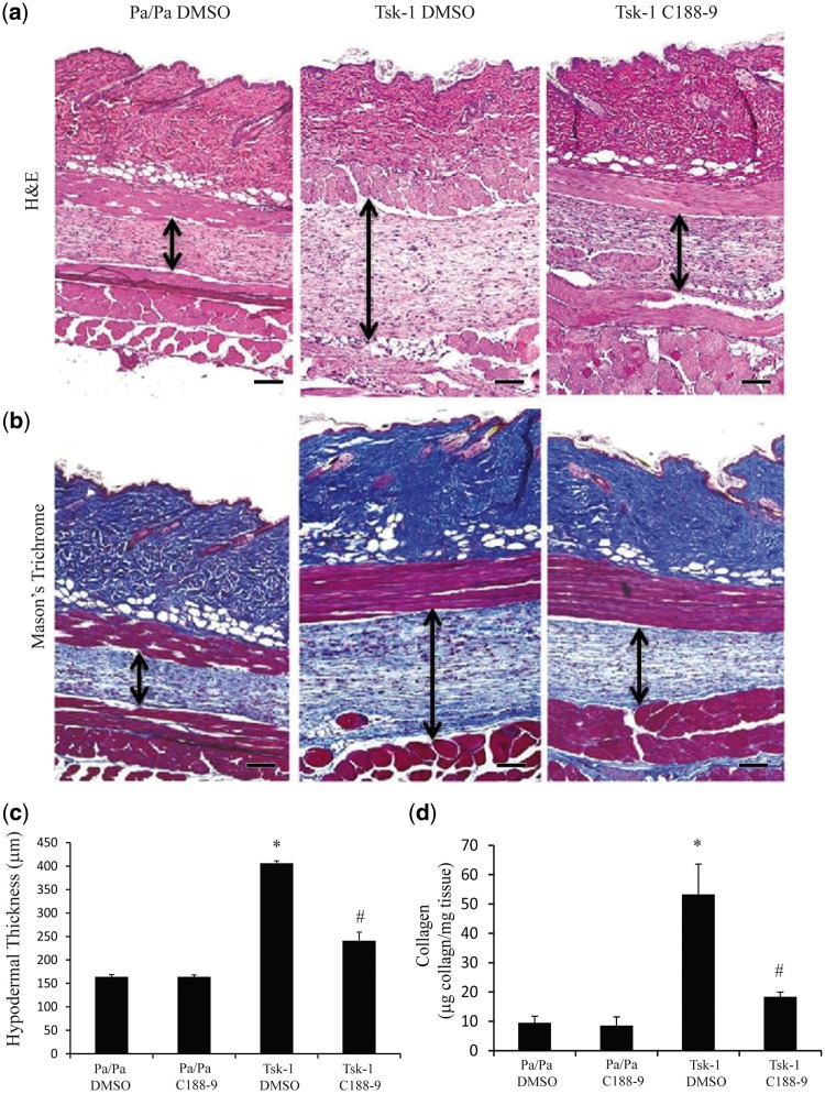
Fig. 4.
STAT3 inhibition with C188-9 decreases hypodermal fibrosis in Tsk-1 mice
Histological examination of skin sections stained with H&E (A) and Mason’s Trichome (B) from Pa/Pa mice treated with DMSO (left; similar results were observed in Pa/Pa mice exposed to C188-9), Tsk-1 mice treated with DMSO (middle) and Tsk-1 mice treated with C188-9 (right). These images revealed that STAT inhibition displayed a reduction in hypodermal fibrosis. Images are representative of 10–12 mice per group. Scale bars: ×10, 100 μm. (C) Hypodermal thickness was measured to determine the degree of fibrosis in the hypodermis section. Data are presented as mean (s.e.m.), n = 10–12 mice per group (*P ≤ 0.05 Pa/Pa DMSO vs Tsk-1 DMSO; **P ≤ 0.05 Tsk-1 DMSO vs Tsk-1 C188-9). (D) Soluble collagen protein levels were measured using Sircol Assay from skin samples obtained from Pa/Pa control mice treated with DMSO or C188-9 and Tsk-1 mice treated with DMSO or C188-9. Data presented as mean microgram collagen per milligram tissue (s.e.m.), n = 10–12 mice per group (*P ≤ 0.05 Pa/Pa DMSO vs Tsk-1 DMSO; **P ≤ 0.05 Tsk-1 DMSO vs Tsk-1 C188-9).

