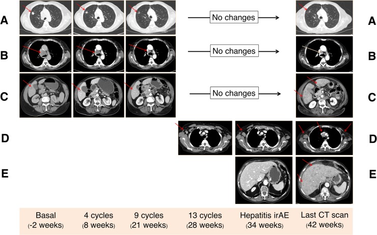
Figure 2. Computed tomography (CT) findings.
Three target lesions (red arrows) were identified in basal CT scan corresponding to lung metastasis (A), mediastinal lymph adenopathy (B), and liver metastasis (C). All these lesions showed a partial response after 4 cycles of nivolumab, which was maintained until the last CT scan (42 weeks). Progression of the axillary right lymph node that appeared as a new lesion at week 28 (D). Flare disease progression in the liver with the apparition of new uncountable liver metastasis in the last CT scan causing hepatic failure (E).

