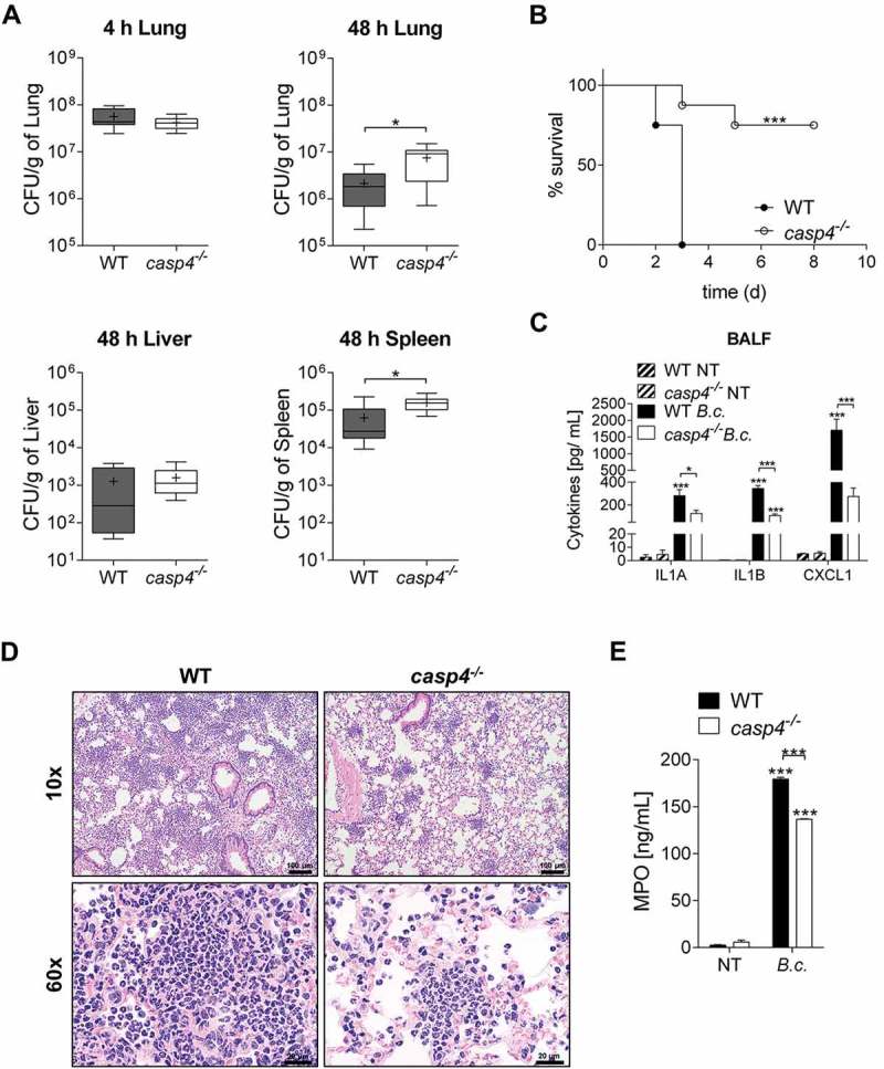
Figure 2.

CASP4/caspase-11 contributes to the restriction of B. cenocepacia in vivo. (a) In vivo CFU from lung, liver and spleen of WT and casp4−/- mice infected with B. cenocepacia K-56 (B.c.) at 4 h (n = 9) and 48 h (n = 10). Pooled data from 2 independent experiments are shown as mean ± SEM. Statistical analysis was performed using two-tailed Student’s t-test. (b) Survival of WT and casp4−/- mice after intratracheal infection with 30 × 106 CFU. Pooled data from 2 independent experiments (n = 8). Statistical analysis was performed using Log-rank (Mantel-Cox) test. (c) Cytokine levels at 24 h in the BALF of WT (n = 7) and casp4−/- (n = 6) mice after intratracheal infection with 30 × 106 CFU. Statistical analysis was performed using two-way ANOVA. (d) Representative 10x (upper panel) and 60x (lower panel) magnification of H&E-stained lung sections from WT and casp4−/- mice showing increased inflammatory infiltrate within the WT sections. (e) MPO levels at 24 h in the BALF of WT and casp4−/- mice treated as in (C). Statistical analysis was performed using two-way ANOVA. *p ≤ 0.05, **p ≤ 0.01, ***p ≤ 0.001.
