
Figure 4.
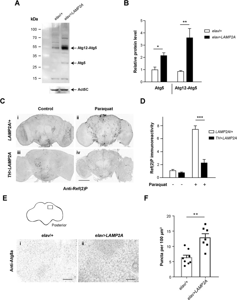
Human LAMP2A enhances macroautophagy in the Drosophila brain. (a, b) Effect of LAMP2A on Atg5 expression. (a) Western blot of head protein extracts from 10-day old control flies (elav/+) and flies expressing human LAMP2A in neurons (elav>LAMP2A) probed with anti-Atg5 antibody. LAMP2A expression markedly increased levels of Atg5 and of the Atg12-Atg5 complex that is required for autophagosome formation. Act5C served as a loading control. (b) Quantification of Atg5 protein and the Atg12-Atg5 complex from 3 independent western blot experiments. (c, d) Effect of LAMP2A on paraquat-induced Ref(2)P accumulation. (c) Anti-Ref(2)P immunostaining in whole-mount adult brains of LAMP2A/+ (panels i and ii) and TH>LAMP2A (panels iii and iv) flies exposed to paraquat (panels ii and iv) or not (panels i and iii). LAMP2A expression prevented paraquat-induced Ref(2)P accumulation (black puncta) suggesting that the human protein is able to maintain efficient autophagic flux under oxidative stress. Scale bar: 100 μm. (d) Quantification of Ref(2)P immunostaining in the central brain region normalized to LAMP2A/+ control not exposed to paraquat (n = 4 or 5 independent brains per condition). (e, f) Effect of LAMP2A on the number of Atg8a-positive puncta. (e) Anti-Atg8a immunostaining in whole-mount adult brains of elav/+ (panel i) and elav>LAMP2A (panel ii) flies. The inset scheme on top shows the posterior neuropil region that was magnified in panels i and ii and in which Atg8a puncta were counted. Scale bars: 10 µm. (f) Quantification of Atg8a-positive dots. Each black circle (elav/+) or square (elav>LAMP2A) represents the score for a different brain. The number of Atg8a puncta that reflect autophagosome formation was markedly increased in LAMP2A-expressing flies. Similar results were obtained in 3 independent experiments.