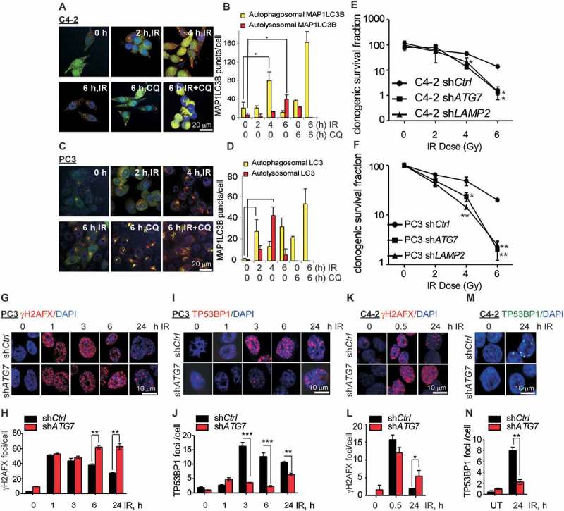
Figure 1.

Inhibition of autophagy leads to an impaired DNA damage response in prostate cancer cells. (a and c) Representative confocal images of C4-2 and PC3 cells, respectively, stably expressing GFP-mCherry-MAP1LC3B at the indicated time following ionizing radiation (IR) ±chloroquine (CQ). (b and d) Quantification of autophagosomal (yellow) and autolysosomal (red) MAP1LC3B from the confocal images of C4-2 and PC3 cells stably expressing GFP-mCherry-MAP1LC3B at the indicated time following IR+/− CQ. (e and f). Clonogenic survival of C4-2 and PC3 cells stably expressing pLKO.1 vector control (shCtrl) or short hairpin RNA for ATG7 (shATG7), or LAMP2 (shLAMP2) following the indicated doses of IR. Representative confocal images and quantification of γH2AFX (g and h) and TP53BP1 (i and j) in PC3 cells stably expressing shCtrl or shATG7 at the indicated times following IR treatment. Nuclei were stained with DAPI. Representative confocal images and quantification of γH2AFX (k and l) and TP53BP1 (m and n) in C4-2 cells stably expressing shCtrl or shATG7 at the indicated times following IR treatment. Nuclei were stained with DAPI. Data shown are the means ± SEM (n = 3) P < 0.05 *. P < 0.01 **. P < 0.001***.
